Life Extension Magazine®

Annual blood testing is the most effective way of detecting imbalances in time to take corrective actions. Countless lives have been spared since people started checking their blood before serious illness develops.
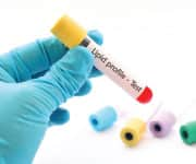
Until recently, the consensus was that people had to fast for 8 to 12 hours prior to having their blood drawn. Before describing studies indicating that fasting may not be required, let’s look at this issue from a practical standpoint.
Most people eat throughout the day and are never in a fasting state. The only exception is the 8-12 hour period prior to a blood test. Results obtained when blood is drawn during this fasting period may not reflect what’s in your blood under normal conditions. This can lead to a false sense of security.
For instance, glucose and triglyceride levels increase after you eat. As it relates to disease risk, how quickly they come down after you finish a meal is important.
Some data suggest that after-meal blood levels of glucose and triglycerides are more accurate predictors of disease risk.1-4
Fasting and non-fasting blood sugar levels impact health and longevity. As you’ll learn in this article, many aging individuals suffer sugar-inflicted damage despite fasting glucose being “normal.”
Triglycerides rise in the blood after a meal and can remain dangerously high for many hours. If one fasts 8-12 hours before a blood draw, triglycerides may appear artificially low compared to where they may be during typical non-fasting periods. This again can create a false sense of security regarding your cardiovascular risk.
Some people neglect having their blood tested because they cannot fast for an 8-12 hour period.
The encouraging news is that for many individuals, a more realistic reading may be obtained when blood is drawn 2-6 hours after a normal meal is consumed.
This article provides a novel rationale for what may become the new optimal time to have your blood drawn.

If you are a participant in a clinical study to assess the effects of a drug on blood fat or sugar levels, it is important for you to not eat for 8-12 hours before your scheduled blood draw.
The reason fasting is important in this instance is to provide researchers with consistent data on the compound they are testing amongst a large study group.
From the standpoint of optimizing your health, you are not part of a study group. Your purpose for having blood tests is to identify hidden risk factors that are correctable before you are stricken with illness or sudden death.
Some credentialed physicians have concluded that individuals do not need to fast before their blood is drawn. They point to studies showing that cholesterol and LDL levels are not significantly altered in most people based on when their blood is drawn (fasting or non-fasting).5-7
In addition, emerging data suggests that after-meal blood levels of triglycerides and glucose may be more important indicators of disease risk than corresponding fasting levels.1-4
What Are Triglycerides?
Like other types of fats, triglycerides are carried in the bloodstream by lipoproteins. Triglyceride-rich lipoproteins contribute to the build-up of plaque in our arteries (atherosclerosis).
The fats we consume in our diet are mainly composed of triglycerides. Therefore, following a fatty meal, blood levels of triglycerides will rise.8
High triglyceride levels increase heart attack and ischemic stroke risk.9,10
After-Meal Fat Damages Arterial Walls

arterial wall.
Postprandial (after-meal) disorders are characterized by fat (and glucose) that persists in the bloodstream many hours after eating. Postprandial disorders are major causes of heart disease and stroke.11,12
As you eat a meal of, say, steak, buttered rolls, and cream in your coffee, fats contained in these foods are absorbed into your circulatory system.
Six hours later, the remnants of your meal should be history. Clearance of fat present in your meal ought to be quick and efficient. After six hours, high levels of absorbed lipids should not be in your bloodstream.
In some people, however, meal remnants (lipoproteins) persist in the blood many hours later. The longer they stick around, the more opportunity they have to trigger growth of atherosclerotic plaque.
Clinical studies show that postprandial triglyceride-rich lipoproteins (like VLDL cholesterol) are powerful instigators of coronary plaque, carotid plaque, and aneurysms of the aorta.11,13,14
Post-meal increases in lipids appear in the blood soon after you begin eating, but usually dissipate by the time your blood is drawn in a “fasting” state (8-12 hours later).
Postprandial disorders are therefore frequently overlooked, and rarely enter into a doctor’s assessment of vascular disease risk. Although their presence may not be apparent from a fasting blood sample, elevated increases in postprandial lipoproteins can be a critically important risk factor for vascular disease.
The danger of after-meal remnants remaining in your blood is one reason why Life Extension® is now suggesting that people consider having their blood drawn 2-6 hours after a normal meal, be it breakfast or lunch. The box on the next page describes some of the mechanisms by which high levels of postprandial lipoproteins damage our arteries.
After-Meal Triglycerides Affect Lipoproteins
Triglycerides are a principal component of postprandial lipoproteins like VLDL.25
Elevated fasting triglycerides can serve as an indirect index of increased postprandial lipo-proteins.26
If fasting triglycerides are high, postprandial lipoproteins are likely to be present. If after-meal triglycerides are high, this is an even stronger indicator of greater postprandial lipoproteins.
Simply put, the higher your triglycerides, the more likely postprandial lipoproteins are also present, potentially putting you at risk for atherosclerotic disease.
However, fasting triglycerides may not be the best way of assessing your atherosclerotic risk. Some people can have fasting triglyceride levels of less than 100 mg/dL, yet still have dangerously high daily levels of postprandial lipoproteins.20
Having your blood drawn in a non-fasting state may provide a better snapshot of what your blood consists of on a normal day.
On blood samples drawn 4-6 hours after a meal, triglycerides over 250 mg/dL indicate a postprandial lipoprotein problem.27,28
A Hidden Cause of Vascular Disease
 |
When lipoproteins like VLDL and chylomicrons linger in the blood for many hours after eating, they are afforded ample opportunity to exert damaging effects on vascular structures. Here are some mechanisms in which high after-meal fats (lipoproteins) accelerate atherosclerosis:
- Postprandial lipoproteins block the artery-relaxing agent known as nitric oxide, while increasing the artery constrictor called endothelin. This induces endothelial dysfunction,12,15 which contributes to the formation of atherosclerotic plaque.16
- Postprandial lipoproteins increase blood levels of cellular adhesion molecules, allowing inflammatory white blood cells to more readily adhere and gain entry to the arterial wall, which also leads to atherosclerotic plaque formation.17
- Postprandial lipoproteins activate blood clotting by increasing factors that both promote blood clotting (thrombosis) and inhibit clot breakdown.18
- Postprandial lipoproteins trigger the formation of a cascade of other abnormal lipoprotein particles that contribute to heart and vascular disease, such as small, dense lipoprotein (small LDL) particles.19
- Postprandial lipoprotein particles insert themselves into atherosclerotic plaque, fueling its growth.20
- Carotid ultrasound studies show that people with elevated postprandial lipoproteins have more carotid plaque than people who do not, independent of their cholesterol values.21,22
- Postprandial lipoproteins predict a greater likelihood of coronary atherosclerotic plaque. People with excessive postprandial (after-meal) abnormalities experience more rapid arterial plaque growth.21-24
FDA’s Absurd Position
We at Life Extension® have argued for the past four decades that optimal triglyceride levels are under 100 mg/dL. The American Heart Association in recent years concurs with our position.
Until a federal judge intervened, the FDA was able to claim that triglyceride levels between 200-499 mg/dL had not been proven to increase vascular disease risk.
I wrote a detailed rebuttal to the FDA’s unscientific position in the May 2016 edition of this publication. Relying on federal government health guidelines can be analogous to living in the medical Dark Ages.
Impact of After-Meal Glucose

We’ve published many articles in Life Extension Magazine® about the lethal impact of high after-meal blood sugar levels.
When blood sugar spikes too high after eating and remains elevated, this presents a significant mortality risk factor.29 These kinds of surges in after-meal glucose (sugar) are associated with prediabetes and diabetes.30,31
Reducing after-meal glucose levels has the potential to help prevent many common aging disorders.
Elevated glucose promotes cardiovascular disease and is associated with an increased risk of dementia, cancer, worse outcomes in cancer patients, and accelerated aging.32-43
Researchers have found that increased two-hour postprandial (after-meal) glucose is an independent risk predictor for cardiovascular and all-cause death.29 During this postprandial period, blood sugar spikes can acutely impair blood flow through vital arteries.44 This can ultimately lead to heart attack or stroke.
After-meal surges in blood sugar directly impair the arteries’ ability to respond to the heart’s demand for an immediate increase in blood flow.12,45
This is one reason that diabetics have such a high prevalence of cardiovascular disorders.46 But even if you don’t have diabetes, a “normal” fasting blood sugar measurement doesn’t protect you against the harmful effects of after-meal glucose spikes.12,45,47,48
People who have normal fasting glucose, but whose glucose levels remain high two-hours after a sugar-laden test drink are diagnosed with “impaired glucose tolerance.” The risk for cardiovascular disease rises sharply in those with impaired glucose tolerance.12,49
One study found that people with impaired glucose tolerance had a 34% higher risk of dying from any form of cardiovascular disease, with a specific 28% greater risk of dying from coronary heart disease.48
Diabetic men with the highest after-lunch blood sugar levels are more than twice as likely to have a cardiovascular event, compared with those with lower levels. In women, that figure rose to a startling 5.5-fold increase.50
In nondiabetic people with metabolic syndrome, every increase in after-meal blood sugar of 18 mg/dL raised the risk of cardiovascular death by 26%.29
These data clearly show that assessing one’s glucose status two or more hours after a normal meal provides critically important information as to one’s underlying disease risk.
Dangers of After-Meal Glucose Not Yet Fully Recognized
 |
When people take blood tests to measure glucose levels, they are asked to fast for 8 to 12 hours.
Doctors request this fasting period because they want a consistent baseline to measure glucose and lipids in comparison with the general population.
The problem with fasting is it can cover up the clinical picture of a person who suffers dangerously high blood sugar many hours following a typical meal.
In other words, a person may artificially drop their fasting glucose to a safe range after fasting 8-12 hours, but in their everyday life, they may never fast for this long.
Thus, a fasting glucose blood test can mask what may be a dangerous postprandial (after-meal) glucose spike.
Even tests that measure long-term glycemic control like hemoglobin A1C (HbA1C) may not adequately detect these post-meal glucose surges.
This means that many individuals are spending at least part of their day in an acute diabetic state. The impact of these multi-hour glucose surges is just now being understood.
To put this in perspective, a study was done where after-meal glucose spikes were impeded using a drug that blocks a digestive enzyme. People taking this drug (acarbose) dropped their heart attack risk by an astounding 91%.62
Cancer and Brain Shrinkage
High-normal blood glucose and elevated insulin increases risk of breast cancer.51-57
Glucose provides fuel for rapidly dividing cancer cells, while insulin promotes tumor growth through multiple pathways.54,58
In a 19-year study, researchers found that participants with impaired fasting glucose of 100 mg/dL or greater had a 49% greater risk of cancer death.59
Those with after-meal glucose above 199 mg/dL had 52% increased cancer death risk. Elevated glucose levels markedly increase an individual’s risk of dying from cancer.59
Glucose levels deemed “high-normal” result in reduced brain volume. A study of 249 volunteers (age early 60s) demonstrated that blood glucose in the high-normal range results in significant brain shrinkage. This shrinkage occurs in regions of the brain involved in memory and other critical functions.60
What may surprise you is that it did not require very “high” glucose levels to cause brain shrinkage. The people in this study classified as having “high” fasting blood sugar levels were below 110 mg/dL. This is the World Health Organization’s threshold for impaired fasting glucose.60 Said differently, glucose levels that mainstream medicine often accept as “normal” are quite hazardous.
In light of evidence linking high normal blood sugar levels with elevated risks of cardiovascular death, cancer, and brain shrinkage, protective steps to reduce glucose levels are critical.61
Those who follow strict calorie restriction diets avoid problems associated with elevated glucose and insulin. For the rest of us, there are steps we can take before meals to impede deadly glucose surges.
What measures you take, however, should begin with a comprehensive blood test panel. Consider not fasting prior to your blood draw. Instead, consume your normal meal/drink and then wait 2-6 hours to have your blood drawn. This will likely enable a more realistic analysis of your underlying disease risks and what can be done to protect your precious health.
What about Homocysteine?
As it relates to the majority of tests, whether your blood is drawn in a fasting or non-fasting state has little impact on the results. This includes sex hormones like DHEA, testosterone, estrogen, and progesterone.
Blood cell counts and assessments of liver and kidney function are unlikely to be significantly affected based on when you ate prior to a blood draw.
When it comes to homocysteine, however, there may be an advantage to having your blood drawn several hours after you eat as opposed to an overnight fast.
The precursor to homocysteine in the blood is the amino acid methionine. Those who ingest red meat often have sharp spikes in homocysteine blood levels.
One study showed that men who consumed large amounts of methionine-rich protein had a steady rise in their homocysteine levels throughout the day, but returned to baseline levels the next morning (after an overnight fast).63
This study has profound implications as it relates to assessing the vascular risk of homocysteine. Numerous studies associate high homocysteine with greater incidence of heart attack64,65 and stroke.66-68 Yet there are inconsistent findings when attempts to lower homocysteine are taken.
A huge overlooked confounding factor is that men who consume a lot of red meat may have elevated homocysteine throughout the day, but it drops to normal the next morning. This would drastically affect the findings of human studies whereby blood draws occur after an overnight fast.
In this case, men who suffered elevated homocysteine almost every day would show normal readings each morning. This would seriously distort the study analysis because men with elevated homocysteine during the day would be placed in the same group whose homocysteine was lower all day. These men with high daily homocysteine are not in safe ranges. They merely measured artificially low because of the overnight fast.
For those who consume a lot of red meat, consider having your blood tested 2-6 hours after the meal while also taking your usual homocysteine-lowering supplements such as folate (5-MTHF form) along with vitamins B2, B6 and B12.
What You Should Do
Standard blood tests are usually done in the fasting state.
Yet a number of studies show that elevated after-meal blood levels of triglycerides and glucose are dangerous.48,69 Ditto for homocysteine that may test normal after an overnight fast, but elevate during a day of high meat ingestion.
Fasting glucose levels alone do not identify individuals with an increased risk of glucose-related disease because they do not detect dangerous after-meal glucose spikes.70,71
The current method of drawing blood only when “fasting” may not adequately measure an individual’s average glucose, triglyceride, homocysteine and postprandial lipoprotein status over the course of a typical day.
By definition, fasting blood tests are conducted eight or more hours after your last meal. This method of only testing blood when in an artificial “fasting” state may not account for vital risk markers specific to you as an individual. In other words, after each meal, your blood sugar and triglycerides will rise, but should return to normal several hours afterwards.
Depending on the consistency and frequency of meals consumed, an individual may silently sustain tissue injuries throughout a typical day that arenot detected when blood is drawn after an 8-12 hour fast.
Conventional dogma is difficult to change, even when common sense and compelling science indicates a better approach.
Based on an accumulating volume of data, consider having your next blood draw in a non-fasting state, as close as possible to what you typically eat and drink on most days (including physical exercise).
Blood Test Super Sale
In 1996, we initiated a low-cost service whereby our members could directly request their own blood tests.
Once a year, we sharply discount the price of our comprehensive Male or Female Blood Test Panels. This enables readers of this magazine to ascertain their disease risk status and initiate preemptive measures before acute illness strikes.
Glucose, homocysteine, and triglycerides are included in these tests, along with a hemoglobin A1C test to help assess long-term glucose control.
As you can readily see on the next page, the large number of tests including sex hormone status provides a comprehensive review of one’s underlying health.
The retail price for these individual tests can be quite high at commercial labs. We offer them for only $199 during the annual Blood Test Super Sale.
As with any purchase, blood tests qualify for Reward Dollars that lower your future cost of supplements.
Upon receiving your order, we immediately send a requisition and list of blood draw stations in your area. You can usually walk in for your blood draw at a time convenient to you. This year we are advising that most people consider having their blood draw done 2-6 hours after a typical meal.
We understand that many of you may want to continue having your blood drawn in a fasting state. The conclusion of a 2016 study showing that fasting is not routinely required prior to lipid testing stated:
“We recommend that non-fasting blood samples be routinely used for the assessment of plasma lipid profiles… non-fasting and fasting measurements should be complementary but not mutually exclusive.”7
Whether you have your blood drawn while fasting or non-fasting, our Wellness Specialists are available to assist you in understanding the results, which come back very quickly nowadays.
To order the Male or Female Blood Test Panel at these discounted prices, call 1-800-208-3444 (24 hours/day).
For longer life,
William Faloon
References
- Langsted A, Freiberg JJ, Nordestgaard BG. Fasting and nonfasting lipid levels: influence of normal food intake on lipids, lipoproteins, apolipoproteins, and cardiovascular risk prediction. Circulation. 2008;118(20):2047-56.
- Onat A, Can G, Cicek G, et al. Fasting, non-fasting glucose and HDL dysfunction in risk of pre-diabetes, diabetes, and coronary disease in non-diabetic adults. Acta Diabetol. 2013;50(4):519-28.
- Moebus S, Gores L, Losch C, et al. Impact of time since last caloric intake on blood glucose levels. Eur J Epidemiol. 2011;26(9):719-28.
- Sacks DB. A1C versus glucose testing: a comparison. Diabetes Care. 2011;34(2):518-23.
- Sidhu D, Naugler C. Fasting time and lipid levels in a community-based population: a cross-sectional study. Arch Intern Med. 2012;172(22):1707-10.
- Doran B, Guo Y, Xu J, et al. Prognostic value of fasting versus nonfasting low-density lipoprotein cholesterol levels on long-term mortality: insight from the National Health and Nutrition Examination Survey III (NHANES-III). Circulation. 2014;130(7):546-53.
- Nordestgaard BG, Langsted A, Mora S, et al. Fasting is not routinely required for determination of a lipid profile: clinical and laboratory implications including flagging at desirable concentration cut-points-a joint consensus statement from the European Atherosclerosis Society and European Federation of Clinical Chemistry and Laboratory Medicine. Eur Heart J. 2016;37(25):1944-58.
- Wojczynski MK, Glasser SP, Oberman A, et al. High-fat meal effect on LDL, HDL, and VLDL particle size and number in the Genetics of Lipid-Lowering Drugs and Diet Network (GOLDN): an interventional study. Lipids Health Dis. 2011;10:181.
- Ninomiya JK, L’Italien G, Criqui MH, et al. Association of the metabolic syndrome with history of myocardial infarction and stroke in the Third National Health and Nutrition Examination Survey. Circulation. 2004;109(1):42-6.
- Antonios N, Angiolillo DJ, Silliman S. Hypertriglyceridemia and ischemic stroke. Eur Neurol. 2008;60(6):269-78.
- Boren J, Matikainen N, Adiels M, et al. Postprandial hypertriglyceridemia as a coronary risk factor. Clin Chim Acta. 2014;431:131-42.
- Ceriello A, Taboga C, Tonutti L, et al. Evidence for an independent and cumulative effect of postprandial hypertriglyceridemia and hyperglycemia on endothelial dysfunction and oxidative stress generation: effects of short- and long-term simvastatin treatment. Circulation. 2002;106(10):1211-8.
- Gronholdt ML, Nordestgaard BG, Nielsen TG, et al. Echolucent carotid artery plaques are associated with elevated levels of fasting and postprandial triglyceride-rich lipoproteins. Stroke. 1996;27(12):2166-72.
- Watt HC, Law MR, Wald NJ, et al. Serum triglyceride: a possible risk factor for ruptured abdominal aortic aneurysm. Int J Epidemiol. 1998;27(6):949-52.
- Jagla A, Schrezenmeir J. Postprandial triglycerides and endothelial function. Exp Clin Endocrinol Diabetes. 2001;109(4):S533-47.
- Maggi FM, Raselli S, Grigore L, et al. Lipoprotein remnants and endothelial dysfunction in the postprandial phase. J Clin Endocrinol Metab. 2004;89(6): 2946-50.
- Ceriello A, Quagliaro L, Piconi L, et al. Effect of postprandial hypertriglyceridemia and hyperglycemia on circulating adhesion molecules and oxidative stress generation and the possible role of simvastatin treatment. Diabetes. 2004;53(3):701-10.
- Silveira A. Postprandial triglycerides and blood coagulation. Exp Clin Endocrinol Diabetes. 2001;109(4):S527-32.
- Ebenbichler CF, Kirchmair R, Egger C, et al. Postprandial state and atherosclerosis. Curr Opin Lipidol. 1995;6(5):286-90.
- Twickler TB, Dallinga-Thie GM, Cohn JS, et al. Elevated remnant-like particle cholesterol concentration: a characteristic feature of the atherogenic lipoprotein phenotype. Circulation. 2004;109(16):1918-25.
- Teno S, Uto Y, Nagashima H, et al. Association of postprandial hypertriglyceridemia and carotid intima-media thickness in patients with type 2 diabetes. Diabetes Care. 2000;23(9):1401-6.
- Mori Y, Itoh Y, Komiya H, et al. Association between postprandial remnant-like particle triglyceride (RLP-TG) levels and carotid intima-media thickness (IMT) in Japanese patients with type 2 diabetes: assessment by meal tolerance tests (MTT). Endocrine. 2005;28(2):157-63.
- Hyson D, Rutledge JC, Berglund L. Postprandial lipemia and cardiovascular disease. Curr Atheroscler Rep. 2003;5(6):437-44.
- Karpe F, Steiner G, Uffelman K, et al. Postprandial lipoproteins and progression of coronary atherosclerosis. Atherosclerosis. 1994;106(1):83-97.
- Nakajima K, Nakano T, Tokita Y, et al. Postprandial lipoprotein metabolism: VLDL vs chylomicrons. Clin Chim Acta. 2011;412(15-16):1306-18.
- Brinton EA, Nanjee MN, Hopkins PN. Triglyceride-rich lipoprotein remnant levels and metabolism: time to adopt these orphan risk factors? J Am Coll Cardiol. 2004;43(12):2233-5.
- Samson CE, Galia AL, Llave KI, et al. Postprandial Peaking and Plateauing of Triglycerides and VLDL in Patients with Underlying Cardiovascular Diseases Despite Treatment. Int J Endocrinol Metab. 2012;10(4):587-93.
- Tiihonen K, Rautonen N, Alhoniemi E, et al. Postprandial triglyceride response in normolipidemic, hyperlipidemic and obese subjects - the influence of polydextrose, a non-digestible carbohydrate. Nutr J. 2015;14:23.
- Lin HJ, Lee BC, Ho YL, et al. Postprandial glucose improves the risk prediction of cardiovascular death beyond the metabolic syndrome in the nondiabetic population. Diabetes Care. 2009;32(9):1721-6.
- Blaak EE, Antoine JM, Benton D, et al. Impact of postprandial glycaemia on health and prevention of disease. Obes Rev. 2012;13(10):923-84.
- Ceriello A, Colagiuri S. International Diabetes Federation guideline for management of postmeal glucose: a review of recommendations. Diabet Med. 2008;25(10):1151-6.
- Barba M, Sperati F, Stranges S, et al. Fasting glucose and treatment outcome in breast and colorectal cancer patients treated with targeted agents: results from a historic cohort. Ann Oncol. 2012;23(7):1838-45.
- Chan JM, Wang F, Holly EA. Sweets, sweetened beverages, and risk of pancreatic cancer in a large population-based case-control study. Cancer Causes Control. 2009;20(6):835-46.
- Coutinho M, Gerstein HC, Wang Y, et al. The relationship between glucose and incident cardiovascular events. A metaregression analysis of published data from 20 studies of 95,783 individuals followed for 12.4 years. Diabetes Care. 1999;22(2):233-40.
- de Vegt F, Dekker JM, Ruhe HG, et al. Hyperglycaemia is associated with all-cause and cardiovascular mortality in the Hoorn population: the Hoorn Study. Diabetologia. 1999;42(8):926-31.
- Hanefeld M, Cagatay M, Petrowitsch T, et al. Acarbose reduces the risk for myocardial infarction in type 2 diabetic patients: meta-analysis of seven long-term studies. Eur Heart J. 2004;25(1):10-6.
- Jee SH, Ohrr H, Sull JW, et al. Fasting serum glucose level and cancer risk in Korean men and women. Jama. 2005;293(2):194-202.
- Lajous M, Willett W, Lazcano-Ponce E, et al. Glycemic load, glycemic index, and the risk of breast cancer among Mexican women. Cancer Causes Control. 2005;16(10):1165-9.
- Michaud DS, Fuchs CS, Liu S, et al. Dietary glycemic load, carbohydrate, sugar, and colorectal cancer risk in men and women. Cancer Epidemiol Biomarkers Prev. 2005;14(1):138-47.
- Monickaraj F, Aravind S, Gokulakrishnan K, et al. Accelerated aging as evidenced by increased telomere shortening and mitochondrial DNA depletion in patients with type 2 diabetes. Mol Cell Biochem. 2012;365(1-2):343-50.
- Ott A, Stolk RP, van Harskamp F, et al. Diabetes mellitus and the risk of dementia: The Rotterdam Study. Neurology. 1999;53(9):1937-42.
- Yaffe K, Blackwell T, Whitmer RA, et al. Glycosylated hemoglobin level and development of mild cognitive impairment or dementia in older women. J Nutr Health Aging. 2006;10(4):293-5.
- Xu W, Qiu C, Winblad B, et al. The effect of borderline diabetes on the risk of dementia and Alzheimer’s disease. Diabetes. 2007;56(1):211-6.
- Nitenberg A, Cosson E, Pham I. Postprandial endothelial dysfunction: role of glucose, lipids and insulin. Diabetes Metab. 2006;32 Spec No2:2s28-33.
- Kawano H, Motoyama T, Hirashima O, et al. Hyperglycemia rapidly suppresses flow-mediated endothelium-dependent vasodilation of brachial artery. J Am Coll Cardiol. 1999;34(1):146-54.
- Reusch JE, Wang CC. Cardiovascular disease in diabetes: where does glucose fit in? J Clin Endocrinol Metab. 2011;96(8):2367-76.
- Title LM, Cummings PM, Giddens K, et al. Oral glucose loading acutely attenuates endothelium-dependent vasodilation in healthy adults without diabetes: an effect prevented by vitamins C and E. J Am Coll Cardiol. 2000;36(7):2185-91.
- Decode Study Group, the European Diabetes Epidemiology Group. Glucose tolerance and cardiovascular mortality: comparison of fasting and 2-hour diagnostic criteria. Arch Intern Med. 2001;161(3):397-405.
- Leiter LA, Ceriello A, Davidson JA, et al. Postprandial glucose regulation: new data and new implications. Clin Ther. 2005;27 Suppl B:S42-56.
- Cavalot F, Petrelli A, Traversa M, et al. Postprandial blood glucose is a stronger predictor of cardiovascular events than fasting blood glucose in type 2 diabetes mellitus, particularly in women: lessons from the San Luigi Gonzaga Diabetes Study. J Clin Endocrinol Metab. 2006;91(3):813-9.
- Contiero P, Berrino F, Tagliabue G, et al. Fasting blood glucose and long-term prognosis of non-metastatic breast cancer: a cohort study. Breast Cancer Res Treat. 2013;138(3):951-9.
- Lawlor DA, Smith GD, Ebrahim S. Hyperinsulinaemia and increased risk of breast cancer: findings from the British Women’s Heart and Health Study. Cancer Causes Control. 2004;15(3):267-75.
- Liao S, Li J, Wei W, et al. Association between diabetes mellitus and breast cancer risk: a meta-analysis of the literature. Asian Pac J Cancer Prev. 2011;12(4):1061-5.
- Muti P, Quattrin T, Grant BJ, et al. Fasting glucose is a risk factor for breast cancer: a prospective study. Cancer Epidemiol Biomarkers Prev. 2002;11(11):1361-8.
- Rapp K, Schroeder J, Klenk J, et al. Fasting blood glucose and cancer risk in a cohort of more than 140,000 adults in Austria. Diabetologia. 2006;49(5):945-52.
- Sieri S, Muti P, Claudia A, et al. Prospective study on the role of glucose metabolism in breast cancer occurrence. Int J Cancer. 2012;130(4):921-9.
- Kabat GC, Kim M, Caan BJ, et al. Repeated measures of serum glucose and insulin in relation to postmenopausal breast cancer. Int J Cancer. 2009;125(11): 2704-10.
- Arcidiacono B, Iiritano S, Nocera A, et al. Insulin resistance and cancer risk: an overview of the pathogenetic mechanisms. Exp Diabetes Res. 2012;2012:789174.
- Hirakawa Y, Ninomiya T, Mukai N, et al. Association between glucose tolerance level and cancer death in a general Japanese population: the Hisayama Study. Am J Epidemiol. 2012;176(10):856-64.
- Cherbuin N, Sachdev P, Anstey KJ. Higher normal fasting plasma glucose is associated with hippocampal atrophy: The PATH Study. Neurology. 2012;79(10):1019-26.
- Kerti L, Witte AV, Winkler A, et al. Higher glucose levels associated with lower memory and reduced hippocampal microstructure. Neurology. 2013;81(20):1746-52.
- Chiasson JL, Josse RG, Gomis R, et al. Acarbose treatment and the risk of cardiovascular disease and hypertension in patients with impaired glucose tolerance: the STOP-NIDDM trial. Jama. 2003;290(4):486-94.
- Verhoef P, van Vliet T, Olthof MR, et al. A high-protein diet increases postprandial but not fasting plasma total homocysteine concentrations: a dietary controlled, crossover trial in healthy volunteers. Am J Clin Nutr. 2005;82(3):553-8.
- Haim M, Tanne D, Goldbourt U, et al. Serum homocysteine and long-term risk of myocardial infarction and sudden death in patients with coronary heart disease. Cardiology. 2007;107(1):52-6.
- Stampfer MJ, Malinow MR, Willett WC, et al. A prospective study of plasma homocyst(e)ine and risk of myocardial infarction in US physicians. Jama. 1992;268(7):877-81.
- Iso H, Moriyama Y, Sato S, et al. Serum total homocysteine concentrations and risk of stroke and its subtypes in Japanese. Circulation. 2004;109(22):2766-72.
- Casas JP, Bautista LE, Smeeth L, et al. Homocysteine and stroke: evidence on a causal link from mendelian randomisation. Lancet. 2005;365(9455):224-32.
- Tanne D, Haim M, Goldbourt U, et al. Prospective study of serum homocysteine and risk of ischemic stroke among patients with preexisting coronary heart disease. Stroke. 2003;34(3):632-6.
- Bansal S, Buring JE, Rifai N, et al. Fasting compared with nonfasting triglycerides and risk of cardiovascular events in women. Jama. 2007;298(3):309-16.
- The DECODE study group on behalf of the Europe an Diabetes Epidemiology Group. Glucose tolerance and mortality: comparison of WHO and American Diabetic Association diagnostic criteria. The Lancet. 1999;354(9179):617-21.
- Nakagami T. Hyperglycaemia and mortality from all causes and from cardiovascular disease in five populations of Asian origin. Diabetologia. 2004;47(3):385-94.

