Life Extension Magazine®
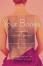
Despite widespread misconception, low bone mass or osteoporosis poses a significant threat to the health and well-being of maturing women and men. A compromised skeletal system not only boosts your risk of life-altering injury; its adverse effects are now known to manifest across multiple systems of the body. Increasingly, cutting-edge researchers are discovering just how critical a role bone health plays in the prevention (or onset) of many killer diseases of aging, from heart disease to diabetes. In this enlightening excerpt from Your Bones, experts Lara Pizzorno, MA, LMT, and Jonathan V. Wright, MD, shed light on the overlooked dangers to healthy bones posed by commonly prescribed pharmaceuticals, surgery, and alcohol. Gastric Bypass: Free Pass? Not for Your BonesGastric bypass (or small-bowel resection) reduces the amount of absorptive surface area in the intestines, and by doing so lessens the body’s ability to absorb not just fat and calories, but also all the nutrients needed to maintain and form healthy bone. The gastric bypass is the leading surgery to treat morbid obesity performed in the United States. Since this operation causes the primary sites where calcium absorption occurs to be bypassed, patients become deficient in calcium and vitamin D. In response to these deficiencies, the body up-regulates the secretion and activity of parathyroid hormone. Parathyroid hormone has two bone-related effects: it causes an increase in the production of the most active form of vitamin D (1,25-dihydroxyvitamin D), which helps us absorb more calcium from food, but it also causes increased bone resorption (bone breakdown) to liberate more calcium for calcium’s many other uses in the body.1,2 Calcium wears a lot of “hats” in the body, playing vital roles in a number of critical physiological processes not related to its use in bone. These include helping blood to clot, so we don’t bleed to death when cut; helping nerves to send impulses and muscles to contract (in the case of the heart muscle, contraction = heartbeat); and regulating our cell membranes, so our cells can allow entry of what they need and send out what they don’t.
Because these activities are essential to life, the body tightly controls the amount of calcium in the blood to ensure that sufficient calcium is available for them. Our bones, where approximately 99% of the calcium in our bodies is stashed, serve as a calcium “bank” from which withdrawals can be made to maintain normal blood concentrations whenever the need arises—which it surely will after gastric bypass (or if we fail to consume calcium-rich foods and/ or supplemental calcium sufficient to meet our body’s needs). Gastric banding, another surgical procedure for morbid obesity, is a safer, potentially reversible, and effective alternative to the Roux-enY gastric bypass that has not been shown to produce as much bone loss as the Roux-en-Y procedure. In gastric banding, an inflatable silicone device is placed around the top portion of the stomach to create a small pouch at the top of the stomach that holds about 3.5 to 6.5 ounces of food. When a person eats, the pouch quickly fills with food, and the band slows its passage from the pouch to the lower part of the stomach. As soon as the upper part of the stomach registers as full, the brain is sent a message that the entire stomach is full, which helps the person eat smaller portions, eat less often, and lose weight over time. Within six to eight years, weight loss from gastric banding is comparable to that achieved by gastric bypass; however, many physicians and patients choose gastric bypass because it results in faster weight loss and resolution of diabetes.3 The fact that some of the weight lost comes from the patient’s bones is somehow overlooked. What Does This Mean for YOU?Either of these surgeries will lessen your body’s ability to absorb calcium and the other nutrients necessary for bone health. If you have had or are considering either of these surgical interventions for morbid obesity, please discuss the potential adverse effects on your bones with your physician. Medical journal articles alerting physicians to these concerns are just beginning to appear, and many doctors remain unaware of these issues.4 Although increasing calcium or vitamin D intake does not suppress parathyroid hormone or prevent the acceleration in bone resorption caused by gastric bypass, it is possible that highly absorbable supplements may help lessen the damage.5 Anyone who has had either of these surgeries should be using calcium supplements. The Liver–Kidney Connection to BoneYou’ve probably heard about how important vitamin D is for bone health. Here’s why: vitamin D stimulates the absorption of calcium from the intestines and also calcium’s resorption from the kidneys, greatly improving the likelihood that adequate calcium will be present in the bloodstream for all the body’s calcium needs.
However, these effects of vitamin D do not occur until after vitamin D has been converted into its most active form in the body. This conversion occurs in two stages, the first of which takes place in the liver, and the second in the kidneys. For this reason, dysfunction in either the liver or the kidneys can compromise vitamin D activation, calcium absorption, and bone health. Approximately 23% of patients with chronic liver disease have osteoporosis. You may be thinking that this couldn’t possibly concern you, that liver disease is uncommon and caused only by alcoholism or hepatitis. You’d be wrong. Today, the most rapidly increasing liver disease is nonalcoholic fatty liver disease or NAFLD, and it is caused by insulin resistance and type 2 diabetes. Following menopause, risk for NAFLD goes up significantly. In a surprising 55% of women over age 60, liver function is compromised by NAFLD.6-9 High blood pressure and diabetes also increase risk for chronic kidney disease, which is estimated to affect 11.5% of adults aged 20 or older in the US.10 What Does This Mean for YOU?NAFLD and other liver diseases often produce no noticeable symptoms and may therefore go undiagnosed. Particularly if you have been diagnosed with metabolic syndrome or type 2 diabetes, be sure your annual physical includes the standard lab tests that check liver function.11 Symptoms of worsening kidney function are also unspecific and may go unnoticed. Symptoms include feeling generally unwell and loss of appetite. Check to be sure that the lab tests run for your annual physical include creatinine. Higher levels of creatinine indicate a decrease in kidney function and the ability to excrete waste products. Anyone suffering from chronic liver or kidney disease is at significantly increased risk for vitamin D deficiency and osteoporosis. Supplemental vitamin D has been found to help lessen bone loss associated with liver and/or kidney disease.12-16 |
What’s Hyperparathyroidism and Why Should My Bones Care?Hyperparathyroidism is overactivity of the parathyroid glands (hyper = excessive, above normal), resulting in excessive production of parathyroid hormone. Hyperparathyroidism is divided into “primary” and “secondary” types. “Primary” hyperparathyroidism is relatively rare. It’s a disease of the parathyroid glands themselves, usually of unknown origin, and is almost always the more severe form. Sometimes surgery is required as part of treatment. “Secondary” hyperparathyroidism is almost always milder, and not always diagnosed as such. Secondary hyperparathyroidism is not a disease (as the primary form is) but a protective response by the body to increase blood levels of calcium from unhealthy low levels caused by inadequate calcium intake or the many other causes listed below. But although parathyroid hormone causes an increase in the body’s production of the most active form of vitamin D (1,25-dihydroxyvitamin D), which helps us absorb more calcium from our intestines, parathyroid hormone also causes increased osteoclast activity and bone resorption (bone breakdown) in order to liberate calcium from bone for calcium’s many other immediate uses in the body. Blood levels of calcium low enough to cause secondary hyperparathyroidism are typically due to not getting enough daily calcium, vitamin D deficiency, chronic kidney disease, chronic liver disease, low levels of stomach acid (hypochlorhydria, relatively common after age 50), malabsorption of calcium (and other minerals, most often caused by “hidden” gluten sensitivity), or gastric bypass surgery. Obesity has also been shown to increase parathyroid hormone levels, which, in addition, tend to increase with age in both men and women. Above normal levels of parathyroid hormone have recently been associated not only with oste oporosis, but also with cognitive decline and senile dementia (Alzheimer’s disease). The connection is most likely explained by the fact that sustained high levels of parathyroid hormone in the brain increase risk of calcium overloading, which leads to impaired blood flow and brain degeneration.17 Fortunately, a recent study involving 37 institutionalized women, ranging in age from their late 70s to late 80s, has shown that consumption of a fortified dairy product, containing only about 17–25% of the recommended daily intakes for calcium and vitamin D, lowered levels of parathyroid hormone and increased levels of both vitamin D and markers of bone formation in just one month.18
What Does This Mean for YOU?
The lab tests ordered at your annual physical should include blood levels of parathyroid hormone. Normal values range from 10–55 picograms per milliliter (pg/mL); however, recent research suggests values higher than 30 may indicate suboptimal intake of calcium and vitamin D.19 Higher than normal levels of parathyroid hormone indicate that you are not meeting your body’s needs for calcium and vitamin D, and that you are at increased risk not only for osteoporosis but cognitive decline and Alzheimer’s disease. Work with your doctor to increase your consumption of calcium and vitamin D, and recheck your levels of parathyroid hormone after a month to two months on your new and improved bone health promotion program. Is Your Thyroid on Overdrive?Hyperthyroidism (a “hyperactive” thyroid) is a well-known risk factor for osteoporosis, regardless of sex or age. The hormones produced and secreted by the thyroid gland regulate the body’s metabolic rate. When thyroid hormone levels are too high, regardless of whether we are pre- or post-menopausal, female or male, it’s like putting the body into overdrive, accelerating all its metabolic activities, including the rate at which bones are remodeled, all the time. Each bone remodeling cycle involves 3-5 weeks of bone breakdown by osteoclasts followed by about 3 months during which osteoblasts lay down new bone to replace the bone that was removed. The result of the fast-forward bone metabolism seen in hyperthyroidism is increased bone resorption that leads to a loss of approximately 10% of bone mass per remodeling cycle. Not surprisingly, this can quickly result in lowered bone mineral density and increased risk of fracture.20,21 Fortunately, hyperthyroidism is relatively uncommon. Gloria Vanderbilt Once Said, “A Woman Can’t Be Too Rich or Too Thin.” She Was Half-Wrong.Anorexia nervosa, an eating disorder characterized by intense fear of gaining weight and becoming fat, despite being underweight (weighing less than 85% of the weight considered normal or healthy for one’s height and build), causes bone loss, particularly in the spine and hip.22,23 This is not surprising since bones cannot be built without a whole team of nutrients and also respond by strengthening when stressed by weight—which is why resistance exercises help build bone. Avoiding food, self-induced vomiting, and use of laxatives, diuretics, and/or appetite suppressants is a sure-fire recipe for bone starvation. Lack of sufficient nourishment not only causes a premenopausal woman to stop menstruating and lose bone, but also causes her to lose muscle and turn into a Skeletor cartoon character look-alike. The stress that muscles put on bone when they contract is a key “time to build more bone” signal. Women are already at a bone-building disadvantage compared to men because our muscles are smaller. Cannibalize your muscles, and you thin your bones. The complete loss of menstrual periods, amenorrhea, occurs largely because the body is no longer willing to use the energy needed to produce estrogen, which regulates osteoclasts, preventing them from removing too much bone. Bone-Busting Patent MedicinesIf you are taking any of the following patent medicines, work with your physician to help compensate for their bone-destroying effects or, if possible, to find an alternative less harmful to your bones. Avandia® (rosiglitazone) and Actos® (pioglitazone): Use of the diabetes patent medicines Avandia® or Actos® for more than a year doubles to triples risk of hip fractures.24-27
These patent medicines, members of a class of patent medicines called thiazolidinediones (also known as glitazones), are insulin-sensitizing medications that account for approximately 21% of the oral blood sugar-lowering patent medicines used in the US. Although their main therapeutic effects occur in fat tissue, muscles, and the liver, studies show they affect bone as well. They do so by triggering mesenchymal stem cells, which can become any one of several different types of cells, including osteoblasts, chondrocytes (cells that produce cartilage), or adipocytes (fat cells), into choosing to become adipocytes. When you take these patent medicines, your body makes more fat and less bone. Because studies have demonstrated accelerated bone loss, impaired bone mineral density, and increased fracture risk for thiazolidinedione users, clinicians have been told to carefully assess the fracture risk in their patients with type 2 diabetes before starting them on thiazolidinediones.28 Anticonvulsants: Barbituates such as phenobarbital or Mysoline® (primidone) alter the metabolism of vitamin D. Dilantin® (phenytoin) interferes with vitamin D and may also cause a deficiency of folate or B6, or a reduction in blood levels of vitamin K, all of which are essential for building and maintaining bone. Chronic Opioid Therapy: Used in the management of chronic pain, opioid patent medicines (e.g., morphine, codeine, hydrocodone, oxycodone, methadone, tramadol) greatly impact the production of a number of hormones, including two with significant effects on bone: estrogen and thyroid-stimulating hormone (TSH). A study of 47 women, aged 30 to 75, who were using oral or transdermal opioids for control of nonmalignant pain found estradiol levels were 57% lower than in control subjects! These patent medicines inhibit estrogen production so effectively that among premenopausal women, menstruation typically ceases soon after initiating opioid therapy. In contrast, opioid patent medicines increase the production of TSH, which directly suppresses bone remodeling. The combined effect of suppressing estrogen production and increasing that of TSH is greatly increased risk of osteoporosis. Women needing opioids for relief of chronic pain should discuss bioidentical hormone replacement with their physicians.29-33 Glucocorticoid patent medicines: Often mistakenly termed “cortisone,” these patent medicines include prednisone, prednisolone, Kenalog®, dexamethasone, and nearly anything else ending in “-one,” along with the nonpatentable Cortef®, which is bioidentical cortisol, but as a prescription often used in excess of normal body levels. These patent medicines kill osteocytes (which is what osteoblasts turn into after they begin secreting the bone matrix). Thus, these patent medicines cause a rapid weakening of bone architecture (within 6 months of initiating treatment) even at very low doses. In addition, the glucocorticoid patent medicines deplete the body of vitamin D3, interfering with normal calcium metabolism and absorption. One reason smoking is so harmful to bone is that nicotine causes the body to produce excess cortisol.34-36 Antacids/Proton-pump inhibitors: For calcium to be absorbed, it must first be made soluble and ionized by stomach acid. These patent medicines inhibit or even totally prevent your body’s ability to produce stomach acid. Are Your Bones Going Up in Smoke?Smokers lose bone more rapidly, have lower bone mass (a full one-third of a standard deviation less at the hip and a one-tenth standard deviation less for all sites combined), and a higher fracture rate. In addition, women who smoke reach menopause, when estrogen levels plummet causing bone loss, up to two years earlier than their nonsmoking peers.37-40 Approximately 19% of the hip fractures occurring in a study that pooled data from three population studies involving a total of 13,393 women and 17,379 men were attributable to smoking.41 In other research, smoking increased risk of spinal osteoporosis in men by 230%!42 More than Two Drinks of Liquor Makes Bone Loss Much QuickerAlcohol has a dose-dependent toxic effect on osteoblast activity. One to two drinks a day appears to be beneficial. More than two drinks a day prevents bone repair and renewal and significantly increases fracture risk.43,44
Using data from the Third National Health and Nutrition Examination Survey, researchers found that moderate drinkers (less than 29 drinks per month) actually had higher BMD than abstainers. Moderate consumption of alcohol translated to 2.1% higher BMD in men and 3.8% higher BMD in postmenopausal women.45 Another large study, this one involving 11,032 women and 5,939 men, found no increase in fracture risk when two ounces or less of alcohol was consumed daily, but drinking more than this increased risk of any osteoporotic fracture by 38% and hip fracture by 68%.46 Your choice of which alcoholic beverage to consume can also affect the health of your bones. Several recent studies suggest moderate intake (no more than two servings a day) of beer and/or wine may have beneficial effects on bone. (One serving of beer = 8 ounces; one serving of wine = 4 ounces.) A study of 1,697 healthy women, of whom 710 were premenopausal, 176 were perimenopausal, and 811 were postmenopausal, found that beer drinkers had slightly higher bone mass.47 A second large study involving 1,182 men, 1,289 postmenopausal woman, and 248 premenopausal women found bone mineral density was 3.0–4.5% greater in men consuming 2 daily drinks of alcohol or beer, and 5.0–8.3 greater in postmenopausal women consuming 1–2 drinks of alcohol or wine daily. More than 2 drinks a day, however, was associated with significantly lower (3.0–5.2% lower) bone mineral density in the hip and spine in men. Beer’s beneficial effects on bone are thought to be due to its silicon content. One can of beer contains around 7 milligrams of silicon; a 4-ounce glass of wine provides around 1 milligram of silicon. (For comparison, a half cup of cooked spinach contains around 5 milligrams of silicon.) Wine’s bone benefits may be linked to its content of phytochemicals, especially the resveratrol present in red wine, which has been shown to have estrogenic effects and might therefore help protect against bone loss in postmenopausal women in whom estrogen levels are low. In rat studies, resveratrol has been shown to have an estrogenic effect and to promote increased BMD in ovariectomized rats (rats whose ovaries have been removed to simulate menopause).48-50 If you have any questions on the scientific content of this article, please call a Life Extension® Wellness Specialistat 1-866-864-3027. | |||||||
| References | |||||||
| 1. Valderas, J. P., S. Velasco, S. Solari, et al. 2009. Increase of bone resorption and the parathyroid hormone in postmenopausal women in the longterm after Roux-en-Y gastric bypass. Obes Surg Aug;19(8):1132–8. Epub 2009 Jun 11. PMID: 19517199. 2. De Prisco, C. and S. N. Levine. 2005. Metabolic bone disease after gastric bypass surgery for obesity. Am J Med Sci Feb;329(2):57–61. PMID: 15711420. 3. Tice, J. A., L. Karliner, J. Walsh, et al. 2008. Gastric banding or bypass? A systematic review comparing the two most popular bariatric procedures. Am J Med Oct;121(10):885–93. PMID: 18823860. 4. Wang, A. and A. Powell. 2009. The effects of obesity surgery on bone metabolism: what orthopedic surgeons need to know. Am J Orthop (Belle Mead NJ) Feb;38(2):77–9. PMID: 19340369. 5. Goode, L. R., R. E. Brolin, H. A. Chowdhury, et al. 2004. Bone and gastric bypass surgery: effects of dietary calcium and vitamin D. Obes Res Jan;12(1):40–7. PMID: 14742841. 6. Nakchbandi, I. A., S. W. van der Merwe. 2009. Current understanding of osteoporosis associated with liver disease. Nat Rev Gastroenterol Hepatol Nov;6(11):660–70. PMID: 19881518. 7. Jean, G., J. C. Terrat, T. Vanel, et al. 2008. Daily oral 25 -hydroxycholecalciferol supplementation for vitamin D deficiency in haemodialysis patients: effects on mineral metabolism and bone markers. Nephrol Dial Transplant Nov;2 3 (11) :3670–6. PMID: 18579534. 8. Jean, G., J. C. Terrat, T. Vanel, et al. 2008. Evidence for persistent vitamin D 1-alpha-hydroxylation in hemodialysis patients: evolution of serum 1,25 -dihydroxycholecalciferol after 6 months of 25-hydroxycholecalciferol treatment. Nephron Clin Pract 110(1):c58–65. PMID: 18724068. 9. Frith, J. and J. L. Newton. 2009. Liver disease in older women. Maturitas Dec 2. [Epub ahead of print] PMID: 19962256. 10. Levey, A. S., L. A. Stevens, C. H. Schmid, et al. 2009. A new equation to estimate glomerular filtration rate. Ann Intern Med May 5;150(9):604– 12. PMID: 19414839. 11. Frith, J., D. Jones and J. L. Newton. 2009. Chronic liver disease in an ageing population. Age Ageing Jan;38(1):11-8. Epub 2008 Nov 22. PMID: 19029099. 12. George, J., H. K. Ganesh, S. Acharya, et al. 2009. Bone mineral density and disorders of mineral metabolism in chronic liver disease. World J Gastroenterol Jul 28;15(28):3516–22. PMID: 19630107. 13. Murlikiewicz, K., A. Zawiasa, M. Nowicki. 2009. [Vitamin D—a panacea in nephrology and beyond] Pol Merkur Lekarski Nov;27(161):437– 41. PMID: 19999813. 14. Moe, S. M., T. Drüeke, N. Lameire, et al. 2007. Chronic kidney disease-mineral-bone disorder: a new paradigm. Adv Chronic Kidney Dis Jan;14(1) :3–12. PMID: 17200038. 15. Spasovski , G. B. 2007. Bone health and vascular calcification relationships in chronic kidney disease. Int Urol Nephrol 39(4):1209–16. Epub 2007 Sep 26. PMID: 17899431. 16. Jean, G., B. Charra and C. Chazot. 2008. Vitamin D deficiency and associated factors in hemodialysis patients. J Ren Nutr Sep;18(5):395–9. PMID: 18721733. 17. Braverman, E. R., T. J. Chen, A. L. Chen, et al. 2009. Age-related increases in parathyroid hormone may be antecedent to both osteoporosis and dementia. BMC Endocr Disord Oct 13;9:21. PMID: 19825157. 18. Bonjour, J. P., V. Benoit, O. Pourchaire , et al. 2009. Inhibition of markers of bone resorption by consumption of vitamin D and calcium-fortified soft plain cheese by institutionalised elderly women. Br J Nutr Oct;102(7):962–6. PMID: 19519975. 19. Braverman, E. R., T. J. Chen, A. L. Chen, et al. 2009. Age-related increases in parathyroid hormone may be antecedent to both osteoporosis and dementia. BMC Endocr Disord Oct 13;9:21. PMID: 19825157. 20. Williams, G. R. 2009. Actions of thyroid hormones in bone. Endokrynol Pol Sep-Oct;60(5):380–8. PMID: 19885809. 21. Zaidi, M., T. F. Davies, A. Zallone, H. C. Blair, et al. 2009. Thyroid-stimulating hormone, thyroid hormones, and bone loss. Curr Osteoporos Rep Jul;7(2):47–52. PMID: 19631028. 22. Legroux-Gérot, I., J. Vignau, M. D’Herbomez, et al. 2007. Evaluation of bone loss and its mechanisms in anorexia nervosa. Calcif Tissue Int Sep;81(3):174–82. PMID: 17668143. 23. Legroux-Gérot, I., J. Vignau, E. Biver, et al. 2010. Anorexia nervosa, osteoporosis and circulating leptin: the missing link. Osteoporos Int Jan 6. [Epub ahead of print] PMID: 20052458. 24. Loke, Y. K., S. Singh, C. D. Furberg. 2009. Longterm use of thiazolidinediones and fractures in type 2 diabetes: a meta-analysis. CMAJ Jan 6;180(1):32–9. PMID: 19073651. 25. Dormuth, C. R., G. Carney, B. Carleton, et al. 2009. Thiazolidinediones and fractures in men and women. Arch Intern Med Aug 10;169(15):1395– 402. PMID: 19667303. 26. Douglas, I. J., S. J. Evans, S. Pocock, et al. 2009. The risk of fractures associated with thiazolidinediones: a self-controlled case-series study. PLoS Med Sep;6(9):e1000154. PMID: 19787025. 27. Meier, C., M. Bodmer, C. R. Meier, et al. 2009. [Thiazolidinediones and skeletal health] Rev Med Suisse Jun 10;5(207):1309–10, 1312–3. PMID: 19626930. 28. McDonough, A. K., R. S. Rosenthal, et al. 2008. The effect of thiazolidinediones on BMD and osteoporosis. Nat Clin Pract Endocrinol Metab Sep;4(9):507–13. PMID: 18695700. 29. Daniell, H. W. 2008. Opioid endocrinopathy in women consuming prescribed sustained-action opioids for control of nonmalignant pain. J Pain Jan;9(1):28–36. PMID: 17936076. 30. Vuong, C., S. H. Van Uum, L. E. O’Dell, et al. 2010. The effects of opioids and opioid analogs on animal and human endocrine systems. Endocr Rev Feb;31(1):98–132. PMID: 19903933. 31. Katz, N., N. A. Mazer. 2009. The impact of opioids on the endocrine system. Clin J Pain Feb;25(2):170–5. PMID: 19333165. 32. Bassett, J. H., P. J. O’Shea, S. Sriskantharajah, et al. 2007. Thyroid hormone excess rather than thyrotropin deficiency induces osteoporosis in hyperthyroidism. Mol Endocrinol May;2 1(5): 1095–107. PMID: 17327419. 33. Zaidi, M., T. F. Davies, A. Zallone, et al. 2009. Thyroid-stimulating hormone, thyroid hormones, and bone loss. Curr Osteoporos Rep Jul;7(2):47– 52. PMID: 19631028. 34. Weng, M.Y., N. E. Lane. 2007. Medication- induced osteoporosis. Curr Osteoporos Rep Dec;5(4):139–45. PMID: 18430387. 35. De Nijs, R. N. 2008. Glucocorticoid-induced osteoporosis: a review on pathophysiology and treatment options. Minerva Med Feb;99(1):23– 43. PMID: 18299694. 36. Silverman, S.L., N. E. Lane. 2009. Glucocorticoid-induced osteoporosis. Curr Osteoporos Rep Mar;7(1):23–6. PMID: 19239826. 37. Slemenda, C.W., S. L. Hui, C. Longcope and C. C. Johnston, Jr. 1989. Cigarette smoking, obesity, and bone mass. J Bone Miner Res Oct;4(5):737– 41. PMID: 2816518. 38. Krall, E. A. and B. Dawson-Hughes. 1991. Smoking and bone loss among postmenopausal women. J Bone Miner Res Apr;6(4):33 1–8. PMID: 1858519. 39. Vestergaard, P. and L. Mosekilde. 2003. Fracture risk associated with smoking: a meta-analysis. J Intern Med Dec;254(6):572–83. PMID: 14641798. 40. Ward, K. D. and R. C. Klesges. 2001. A meta-analysis of the effects of cigarette smoking on bone mineral density. Calcif Tissue Int May;68(5) :259– 70. PMID: 11683532. 41. Høidrup, S., E. Prescott, T. I. Sørensen, et al. 2000. Tobacco smoking and risk of hip fracture in men and women. Int J Epidemiol Apr;29(2):253– 9. PMID: 10817121. 42. Seeman, E., L. J. Melton, 3rd, W. M. O’Fallon, et al. 1983. Risk factors for spinal osteoporosis in men. Am J Med Dec;75(6):977–83. PMID: 6650552. 43. Chakkalakal, D. A. 2005. Alcohol-induced bone loss and deficient bone repair. Alcohol Clin Exp Res Dec;29(12):2077–90. PMID: 16385177. 44. Broulik, P. D., J. Rosenkrancová, P. Růžička, et al. 2009. The effect of chronic alcohol administration on bone mineral content and bone strength in male rats. Physiol Res Nov 20. [Epub ahead of print] PMID: 19929136. 45. Wosje, K. S. and H. J. Kalkwarf. 2007. Bone density in relation to alcohol intake among men and women in the United States. Osteoporos Int Mar;18(3):391–400. PMID: 17091218. 46. Kanis, J. A., H. Johansson, O. Johnell, et al. 2005. Alcohol intake as a risk factor for fracture. Osteoporos Int Jul;16(7):737–42. PMID: 15455194. 47. Pedrera-Zamorano, J. D., J. M. Lavado-Garcia, R. Roncero-Martin, et al. 2009. Effect of beer drinking on ultrasound bone mass in women. Nutrition Oct;25(10):1057–63. Epub 2009 Jun 13. PMID: 19527924. 48. Tucker, K. L., 2009. Jugdaohsingh R, Powell JJ, et al. Effects of beer, wine, and liquor intakes on bone mineral density in older men and women. Am J Clin Nutr Apr;89(4):1188–96. Epub 2009 Feb 25. PMID: 19244365. 49. Liu, Z. P., W. X. Li, B. Yu, et al. 2005. Effects of trans-resveratrol from Polygonum cuspidatum on bone loss using the ovariectomized rat model. J Med Food Spring 8(1):14-9. PMID: 15857203. 50. King, R. E., J. A. Bomser and D. B. Min. Bioactivity of resveratrol. Compr Rev Food Sci Food Saf 5:65–70. DOI 10.1111/j.1541-4337.2006.00001.x , http:// www3.interscience.wiley.com/journal/118607162/ abstract. Excerpted from Your Bones, How You Can Prevent Osteoporosis & Have Strong Bones for Life – Naturally by Lara Pizzorno, MA, LMT with Jonathan V. Wright, MD. Reprinted with permission from Praktikos Books. |


