Life Extension Magazine®
As scientists unravel the secrets of longevity, they are increasingly focusing their studies on centenarians (people who have lived to be over 100 years old). This field of inquiry has revealed deep insights into the protective factors that enable this growing population to live long, healthy lives.1 A series of studies reveals how aging humans can pro-actively “turn on” protective factors that may help promote long, disease-free life. Remarkable insights from an Italian research group, together with late-breaking news from medical researchers in the US suggest that a targeted approach to reducing inflammation and optimizing cardiovascular risk factors may dramatically impact human longevity.1-4 Survivors of InflammagingLed by Dr. Claudio Franceschi, Italian researchers studying centenarians have learned that they represent, in some ways, a special biological group—they appear to be genetically more resistant to inflammation and its devastating consequences.5 We have known for decades that inflammation and oxidation participate in a devious and destructive interplay that underlies most of the changes that have long been thought to be inevitable consequences of aging.6,7 As a result of his work with humans of very advanced age and their unique ability to suppress inflammation, Dr. Franceschi coined the term inflammaging to summarize his team’s epiphany: as we age, our acquired immunity wanes, leaving us at growing risk for infections and cancers, while our innate immunity is largely preserved, putting us at steadily increasing risk for the ravages of inflammation.1 According to Dr. Franceschi, “Inflammaging is considered the common and most important driving force of age-related pathologies, such as neurodegeneration, atherosclerosis, diabetes, and sarcopenia [loss of muscle mass], among others, all of which share an inflammatory cause.”
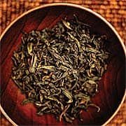
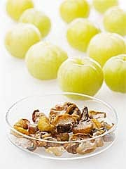
Since Dr. Franceschi’s seminal 2003 work, researchers from around the world have embraced and expanded on the inflammaging concept, bringing it increasingly to the attention of the general public and even conservative thinkers in the medical mainstream.5,8-13 The culmination of this work was published recently in late 2008, when the New York Times splashed the headline “Cholesterol-Fighting Drugs Show Wider Benefit.”14 The occasion was the publication of the dramatic results of JUPITER, a study of 17,802 apparently healthy men and women with normal low-density lipoprotein (LDL) levels but high levels of the inflammatory marker protein called C-reactive protein, or CRP.4 This study demonstrated that people without abnormal lipid profiles, but with signs of increased inflammation had remarkably lower rates of cardiovascular events such as heart attack, stroke, and their consequences, when they took a “statin” drug called rosuvastatin (Crestor®). Rosuva-statin reduced LDL levels by 50% and CRP levels by 37%. In this study, the reduction of inflammation seem-ed to be critical to avoiding a cardiovascular event. In fact, the effects were so powerfully positive that the study was halted after an average follow-up period of just under two years!4 The aftershocks in the medical community continue to reverberate, as physicians are grappling to understand this powerful evidence of the impact of inflammation on cardiovascular disease. While statins form an important part of our armamentarium against cardiovascular disease, they sometimes carry adverse effects.15 Fortunately, two nutrients may produce similar benefits without concern of side effects. To reduce destructive inflammatory reactions, an extract from black tea called theaflavins has demonstrated powerful antioxidant effects and a remarkable ability to control inflammation at the genetic level.3 To help regulate blood lipid levels, Indian gooseberry or amla has produced exciting results in human clinical trials with the added benefit of reducing oxidative damage to fats that can lead to early atherosclerotic changes.2 Theaflavins and NutrigenomicsMost people by now are aware of the astonishing powers of green tea and its antioxidant polyphenolic flavonoid components such as epigallocatechin gallate (EGCG).16 Now, a related family of compounds found in black tea, called theaflavins, is attracting attention for its promising effects with regard to aging and longevity.17 The theaflavins are at the forefront of the exciting new field of nutrigenomics—the study of nutritional molecules that can directly influence the expression of genes, that is, how individual genes switch themselves on and off to regulate basic biological processes.18-22 Since inflammatory processes are controlled by genes and the proteins they produce, our ability to control those genes opens a vast new field for effective interventions that will control inflammation and hence the inflammatory consequences of aging, or inflammaging. Well-known nutrigenomics molecules include, for example, the omega-3 fatty acids which regulate lipid profiles,23 insulin responses,24 and inflam-matory mediators,25 and the spice-derived curcumin molecule that regulates transcription factors involved in inflammation and cancer induction.26 These black tea components (theaflavins) now join this impressive group of molecules.
Given the LDL-lowering effect of drinking multiple cups of tea per day as shown in epidemiological studies, cardiologists at Vanderbilt University recently studied the impact of a theaflavin-enriched green tea extract on lipid profiles of subjects with mild and moderately elevated cholesterol.27 Studying 240 men and women in China, the researchers randomly assigned patients to receive either placebo or a theaflavin-enriched green tea extract (comprising 75 mg theaflavins, 150 mg of green tea catechins, and 150 mg other tea polyphenols) daily for 12 weeks. At the end of the study, the theaflavin-supplemented patients experienced decreases in their total cholesterol and LDL by 11% and 16% respectively, while placebo recipients had no change at all. No significant adverse effects were observed, and the researchers concluded that this theaflavin-enriched extract was “an effective adjunct to a low-saturated fat diet.” Further evidence for black tea was provided by Boston cardiologists in 2001, when they studied the impact of supplementation on endothelial dysfunction. The researchers randomly assigned 66 patients with coronary artery disease to consume either black tea or water; the study was designed in a “cross-over” fashion so that all subjects got both the tea and the water at different times (this allows comparisons within individuals as well as between subjects). The findings were dramatic: both short- and long-term tea consumption significantly improved blood flow in arteries, as detected by ultrasound measurements; this flow is controlled by endothelial cells, and is typically decreased in patients with cardiovascular diseases. The researchers concluded that “short- and long-term black tea consumption reverses endothelial vasomotor dysfunction in patients with coronary artery disease.”28
The novel anti-inflammatory characteristics of black tea-derived theaflavin were described in a 2004 paper.3 These researchers were specifically interested in the gene that produces the inflammatory cytokine interleukin-8 (IL-8), which is responsible for much of the acute inflammation seen in conditions such as asthma, gum disease, inflammatory bowel disease, and perhaps even cancer and cardiovascular disease.29-31 Remarkably, these researchers found that theaflavin inhibited IL-8 production. Even more remarkably, the effect was traced to theaflavin’s ability to inhibit the transcription of the IL-8 gene—in other words, theaflavin blocked the gene from actually expressing its product, the inflammatory cytokine.3 This pinpoint accuracy opens the door to many specific applications of theaflavin as an anti-inflammatory nutrigenomic agent. British researchers found that black tea consumption reduces the rate of platelet aggregation.32 The aggregation of platelets contributes to clot formation, which can precipitate deadly cardiovascular events. In 2004 Japanese researchers were able to pin down black tea’s platelet anti-aggregating effect to a small group of highly active concentrated theaflavins.33 The powerful anti-inflammatory, anticancer, and longevity-inducing qualities of highly active theaflavin fractions has resulted in publication of several dozen laboratory studies, each providing more high-resolution details about these molecules.34-38
A compelling human trial drives home the incredible potential of theaflavin extracts at fitting into the inflammaging model of human disease by reducing inflammatory processes across a wide spectrum of actions. In this 2007 study of 12 human volunteers, eight were supplemented with the highly purified extract for a week, while four received placebo. At the end of the week, the subjects received injections of one of the most powerful stimulators of inflammation known to science: a bacterial cell membrane component called lipopolysaccharide (LPS). LPS in modest doses can induce shock, coma, and even death, so naturally these were only minute and safe doses, but clearly the volunteers were expected to show some evidence of acute inflammatory reactions. In addition to clinical monitoring, the investigators drew blood samples to monitor for early signs of inflammation, particularly those involving “inducible” cytokines such as TNF-alpha, IL-6, IL-8, and CRP.39,40 Astonishingly, the supplemented subjects had a 56% reduction in levels of these cytokines even before they received the inflammatory challenge! Equally importantly, supplemented subjects experienced a 52% increase in levels of the protective, anti-inflammatory cytokine called IL-10, which is involved in prevention of viral respiratory infections, for example.41 The supplemented patients also demonstrated lower rates of production of the inflammation-generating transcription factor NF-kB (71%), the cytokine-generating enzyme COX-2 (72%), and the adhesion molecule ICAM-1.40 C-reactive protein, or CRP, rose dramatically as expected in the placebo recipients following the inflammatory challenge. Remarkably, though, that elevation was 75% greater in the placebo group than in the supplemented group. Since we know that reducing CRP levels can be life saving,4 this is direct evidence of the benefits of this concentrated theaflavin preparation!42,43 AmlaThe Indian gooseberry (Emblica officinalis) has been well known to practitioners of Ayurvedic medicine (native to India) for more than 3,000 years.44 Long-ignored by Western scientists, the Indian gooseberry, also known as amla, belongs to a group of herbal preparations that, according to historic texts, “promote longevity and induce nourishment.”45 We are finally catching up with our past as evidence accumulates that the amla berry functions at several different levels to slash cardiovascular risk factors. Pure extracts of amla have now been shown to act in precise ways to break the cycle of oxidation, inflammation, and plaque formation. Amla’s powerful antioxidant properties, first recognized in modern terms in 1936,46 seem to underlie most of its beneficial effects (though there appear to be other effects as well). Indian researchers in 1999 showed that the “tannoid principles” of amla are the chief antioxidant compounds in the fruit.47 Amla extracts were in fact ranked by German researchers among the most active botanical agents at preventing lipid peroxidation, the oxidation of fats that triggers the cascade of atherosclerosis and many other age-related conditions.48 Japanese researchers have shown that amla extract administration can reverse many age-related changes in the kidneys of rats,49 a vital finding because kidney function typically deteriorates with age as a result of accumulation of advanced glycation end products (AGEs) and collagen cross linking in the cells and tissues.50,51 Indian researchers studying the effects of amla on ischemic heart disease in rats found that they could completely prevent the “ischemia-reperfusion injury” to heart muscle that occurs when injured cardiac cells are re-supplied with oxygen-rich blood.52 Much of the excitement being generated about cardioprotection in scientific circles today53 is related to the fact that amla extracts not only reduce the oxidation of fat molecules in cell membranes and in blood—they actually reduce levels of dangerous fats while increasing levels of beneficial high-density lipoprotein (HDL). Amla extracts inhibit LDL oxidation (a critical first step in atherosclerosis) more powerfully than the antioxidant drug probucol!54 In an animal model in which rats were fed high-cholesterol diets, amla extracts significantly lowered total cholesterol and LDL levels. The excitement of the researchers is palpable as they conclude that “amla may be effective for hypercholesterolemia [elevated total cholesterol] and the prevention of atherosclerosis.” The mechanisms behind these effects are similar to those of modern cholesterol-lowering prescription drugs.55 For example, amla extracts inhibit the enzyme system called HMG-CoA reductase, which is responsible for production of cholesterol in the liver,53 thereby lowering serum cholesterol levels. Amla also enhances degradation and elimination of cholesterol in animal models,53 as do drugs such as the fibrates (gemfibrozil, clofibrate, and others).56,57 Finally, as elegantly demonstrated by Chinese researchers, amla extracts beneficially affect the endothelial linings of blood vessels, preventing inflammatory cells from “sticking” to them in the first steps of atherosclerosis, and preventing the overgrowth of smooth muscle cells that is the next step in disease production.58-60 In short, concentrated amla extracts work in not one but at least three distinct and complementary ways to reduce cardiovascular risk. Human trials with amla are nothing short of remarkable. Food scientists in New Delhi studied a group of men aged 35-55 years, with either normal or elevated cholesterol levels,61 supplementing them with amla extracts. The subjects received the supplement for 28 consecutive days. Men with both normal and elevated cholesterol levels showed a decrease in total cholesterol levels, and both groups rose right back to nearly their original levels two weeks after stopping the supplement. A more detailed study was reported by physiologists working at the All India Institute of Medical Sciences in New Delhi in 2001.2 These researchers used amla in the form of a traditional Indian supplement rich in vitamin C called “chyawanprash.” Ten healthy young men supplemented their diets with either chyawanprash or vitamin C alone daily for eight weeks, and then no supplement for the subsequent eight weeks. Lipid profiles and glucose tolerance tests were done before supplementation and at four, eight, 12, and 16 weeks. The results were remarkable for such a small study: compared with the vitamin C-only group after eight weeks, the group receiving the amla-containing supplement experienced a nearly 11% increase in HDL levels, more than a 16% drop in LDL levels, and a drop in the LDL:HDL cholesterol ratio of more than 33%.2 These numbers are actually equal to or better than similar measurements in a recent study of the fibrate class of prescription drugs, in which significant safety concerns were raised!57 Subjects in the supplemented group also experienced a reduction of nearly 14% in fasting blood glucose and other measures of glucose tolerance—another beneficial effect for vascular health. Still more recent studies have been conducted on a highly concentrated and purified form of the most active tannins and polyphenols from the amla berry called Amlamax.®62,63 Not all of these studies have been fully reviewed by a panel of experts (the “peer review” process), but the results they report are impressive and deserve mention. In the most recent and compelling study, 25 subjects took the amla preparation (500 mg/day) for three months, during which time 27 took placebo. Twelve vital parameters related to cardiovascular health were measured before the study and then monthly during the study. Remarkably, ten of these parameters showed substantial improvement in the supplemented group! Here is a summary of the findings.63 Lipid Profiles: Subjects who took the concentrated amla preparation experienced a significant 18% increase in their HDL levels compared with placebo recipients. Higher HDL levels are strongly associated with positive cardiovascular protection. That’s because HDL removes cholesterol from the arterial wall (via the reverse cholesterol transport process) and transports it back to the liver for excretion.64 Supplemented subjects also tended towards a reduction in levels of the dangerous blood lipid known as very low-density lipoprotein (VLDL). Triglycerides are actual fat components found in the various lipoprotein molecules, and they contribute independently to cardiovascular risk. Subjects supplemented with the amla extract tended towards lower levels of triglycerides compared with placebo recipients.63 Paraoxonase-1: This enzyme, also known as PON-1, is a vital antioxidant that forms part of the beneficial HDL complex, and contributes directly to its anti-atherosclerotic activity. People with higher levels of PON-1 have a lower risk of cardiovascular outcomes.65 The PON-1 enzyme acts by reducing the dangerous oxidation of LDL fats—one of the early steps in cardiovascular disease. Amla-supplemented subjects had an impressive increase in their PON-1 activity compared with placebo recipients.63
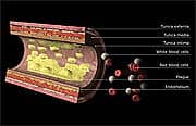
TBARS: This substance (thiobarbituric acid reducing substance) is produced when fat molecules undergo oxidative damage, so TBARS is a direct measure of how much oxidative damage has actually taken place.66 Supplemented patients had a whopping 52% reduction in their TBARS levels after three months of treatment, compared with controls! This finding is stunning direct evidence of the powerful oxidation-protective effect of the special amla concentrate.63 Oxidized LDL: Low-density lipoproteins are among the most dangerous elements when it comes to lipid profiles, in large part because the fats carried by these complexes are especially vulnerable to oxidation, which is among the major triggers of the endothelial damage that presages atherosclerosis.67 Like TBARS, measurements of oxidized LDL are direct indicators of how much actual fat oxidation is taking place, and hence give a “real-time” estimate of one’s cardiovascular risk level.68 In the amla-concentrate supplementation study, supplemented patients experienced a 17% reduction in their oxidized LDL levels, compared with only an 8% reduction in placebo patients—a significant difference that again provides direct proof of this amla preparation’s cardioprotective effects.63 Sialic Acid: This molecule, a ubiquitous component of specialized molecules called glycoproteins, has recently been found to be a sensitive predictor of cardiovascular risk when measured in blood.69,70 A very dramatic and significant drop in serum sialic acid levels was shown among amla-supplemented patients compared with controls.63 SummaryA myriad of published studies have thrust the concept of inflammaging front and center into the scientific community’s awareness. While Life Extension members were long ago warned about the dangers of chronic inflammation and oxidation, large and powerful studies methodically document the benefit of treating elevated CRP and abnormal lipid levels before heart attack, stroke, and other common diseases are clinically manifested.4 The amla berry has been shown to lower cardiovascular risk through its beneficial effects on lipid profiles and by interfering with the way fat molecules undergo deadly oxidation reactions that contribute to chronic inflammation.
When we add the remarkable nutrigenomic effects of theaflavins, which directly control the genes that modulate inflammatory processes, we find a potent “one-two punch” in defeating the deadly forces of inflammaging. Our goal is to use these targeted, scientifically validated supplements to recreate for everyone the centenarians’ proven resistance to the inflammaging process. Our longer-term goal is to push the frontiers of human aging so far that a centenarian is no longer a matter of curiosity, as we all experience the extended good health that intelligent use of technology will provide. If you have any questions on the scientific content of this article, please call a Life Extension Health Advisor at 1-800-226-2370. | ||||||||||||
| References | ||||||||||||
| 1. Franceschi C, Bonafe M. Centenarians as a model for healthy aging. Biochem Soc Trans. 2003 Apr;31(2):457-61. 2. Manjunatha S, Jaryal AK, Bijlani RL, Sachdeva U, Gupta SK. Effect of chyawanprash and vitamin C on glucose tolerance and lipoprotein profile. Indian J Physiol Pharmacol. 2001 Jan;45(1):71-9. 3. Aneja R, Odoms K, Denenberg AG, Wong HR. Theaflavin, a black tea extract, is a novel anti-inflammatory compound. Crit Care Med. 2004 Oct;32(10):2097-103. 4. Ridker PM, Danielson E, Fonseca FA, et al. Rosuvastatin to prevent vascular events in men and women with elevated C-reactive protein. N Engl J Med. 2008 Nov 9. 5. Franceschi C. Inflammaging as a major characteristic of old people: can it be prevented or cured? Nutr Rev. 2007 Dec;65(12 Pt 2):S173-6. 6. Mach F. Inflammation is a crucial feature of atherosclerosis and a potential target to reduce cardiovascular events. Handb Exp Pharmacol. 2005;(170):697-722. 7. Fillit HM, Butler RN, O’Connell AW, et al. Achieving and maintaining cognitive vitality with aging. Mayo Clin Proc. 2002 Jul;77(7):681-96. 8. Mishto M, Santoro A, Bellavista E, et al. Immunoproteasomes and immunosenescence. Ageing Res Rev. 2003 Oct;2(4):419-32. 9. Giunta S. Is inflammaging an auto[innate]immunity subclinical syndrome? Immun Ageing. 2006;312. 10. Franceschi C, Capri M, Monti D, et al. Inflammaging and anti-inflammaging: a systemic perspective on aging and longevity emerged from studies in humans. Mech Ageing Dev. 2007 Jan;128(1):92-105. 11. Florez H, Troen BR. Fat and inflammaging: a dual path to unfitness in elderly people? J Am Geriatr Soc. 2008 Mar;56(3):558-60. 12. Forte GI, Cala C, Scola L, et al. Role of environmental and genetic factor interaction in age-related disease development: the gastric cancer paradigm. Rejuvenation Res. 2008 Apr;11(2):509-12. 13. Katepalli MP, Adams AA, Lear TL, Horohov DW. The effect of age and telomere length on immune function in the horse. Dev Comp Immunol. 2008;32(12):1409-15. 14. Belluck P. Cholesterol-fighting drugs show wider benefit. The New York Times. November 10, 2008;A1. 15. Escobar C, Echarri R, Barrios V. Relative safety profiles of high dose statin regimens. Vasc Health Risk Manag. 2008;4(3):525-33. 16. Beltz LA, Bayer DK, Moss AL, Simet IM. Mechanisms of cancer prevention by green and black tea polyphenols. Anticancer Agents Med Chem. 2006 Sep;6(5):389-406. 17. Cameron AR, Anton S, Melville L, et al. Black tea polyphenols mimic insulin/insulin-like growth factor-1 signalling to the longevity factor FOXO1a. Aging Cell. 2008 Jan;7(1):69-77. 18. Fernandes G. Progress in nutritional immunology. Immunol Res. 2008;40(3):244-61. 19. Kussmann M, Blum S. OMICS-derived targets for inflammatory gut disorders: opportunities for the development of nutrition related biomarkers. Endocr Metab Immune Disord Drug Targets. 2007 Dec;7(4):271-87. 20. Caramia G. Omega-3: from cod-liver oil to nutrigenomics. Minerva Pediatr. 2008 Aug;60(4):443-55. 21. Crujeiras AB, Parra D, Milagro FI, et al. Differential expression of oxidative stress and inflammation related genes in peripheral blood mononuclear cells in response to a low-calorie diet: a nutrigenomics study. OMICS. 2008 Aug 7. 22. Gillies PJ. Preemptive nutrition of pro-inflammatory states: a nutrigenomic model. Nutr Rev. 2007 Dec;65(12 Pt 2):S217-20. 23. Lindi V, Schwab U, Louheranta A, et al. Impact of the Pro12Ala polymorphism of the PPAR-gamma2 gene on serum triacylglycerol response to n-3 fatty acid supplementation. Mol Genet Metab. 2003 May;79(1):52-60. 24. Tsitouras PD, Gucciardo F, Salbe AD, Heward C, Harman SM. High omega-3 fat intake improves insulin sensitivity and reduces CRP and IL6, but does not affect other endocrine axes in healthy older adults. Horm Metab Res. 2008 Mar;40(3):199-205. 25. Blazovics A, Hagymasi K, Pronai L. Cytokines, prostaglandins, nutritive and non-nuitritive factors in inflammatory bowel diseases. Orv Hetil. 2004 Dec 12;145(50):2523-9. 26. Chen HW, Lee JY, Huang JY, et al. Curcumin inhibits lung cancer cell invasion and metastasis through the tumor suppressor HLJ1. Cancer Res. 2008 Sep 15;68(18):7428-38. 27. Maron DJ, Lu GP, Cai NS, et al. Cholesterol-lowering effect of a theaflavin-enriched green tea extract: a randomized controlled trial. Arch Intern Med. 2003 Jun 23;163(12):1448-53. 28. Duffy SJ, Keaney JF, Jr., Holbrook M, et al. Short- and long-term black tea consumption reverses endothelial dysfunction in patients with coronary artery disease. Circulation. 2001 Jul 10;104(2):151-6. 29. Kebschull M, Demmer R, Behle JH, et al. Granulocyte chemotactic protein 2 (gcp-2/cxcl6) complements interleukin-8 in periodontal disease. J Periodontal Res. 2008 Oct 7. 30. Higashimoto Y, Yamagata Y, Taya S, et al. Systemic inflammation in COPD and asthma: similarities and differences. Nihon Kokyuki Gakkai Zasshi. 2008 Jun;46(6):443-7. 31. Aukrust P, Yndestad A, Smith C, Ueland T, Gullestad L, Damas JK. Chemokines in cardiovascular risk prediction. Thromb Haemost. 2007 May;97(5):748-54. 32. Steptoe A, Gibson EL, Vuononvirta R, et al. The effects of chronic tea intake on platelet activation and inflammation: a double-blind placebo controlled trial. Atherosclerosis. 2007 Aug;193(2):277-82. 33. Sugatani J, Fukazawa N, Ujihara K, et al. Tea polyphenols inhibit acetyl-CoA:1-alkyl-sn-glycero-3-phosphocholine acetyltransferase (a key enzyme in platelet-activating factor biosynthesis) and platelet-activating factor-induced platelet aggregation. Int Arch Allergy Immunol. 2004 May;134(1):17-28. 34. Cai F, Li CR, Wu JL, et al. Theaflavin ameliorates cerebral ischemia-reperfusion injury in rats through its anti-inflammatory effect and modulation of STAT-1. Mediators Inflamm. 2006;2006(5):30490. 35. Siddiqui IA, Adhami VM, Afaq F, Ahmad N, Mukhtar H. Modulation of phosphatidylinositol-3-kinase/protein kinase B- and mitogen-activated protein kinase-pathways by tea polyphenols in human prostate cancer cells. J Cell Biochem. 2004 Feb 1;91(2):232-42. 36. Siddiqui IA, Raisuddin S, Shukla Y. Protective effects of black tea extract on testosterone induced oxidative damage in prostate. Cancer Lett. 2005 Sep 28;227(2):125-32. 37. Siddiqui IA, Zaman N, Aziz MH, et al. Inhibition of CWR22Rnu1 tumor growth and PSA secretion in athymic nude mice by green and black teas. Carcinogenesis. 2006 Apr;27(4):833-9. 38. Zykova TA, Zhang Y, Zhu F, Bode AM, Dong Z. The signal transduction networks required for phosphorylation of STAT1 at Ser727 in mouse epidermal JB6 cells in the UVB response and inhibitory mechanisms of tea polyphenols. Carcinogenesis. 2005 Feb;26(2):331-42. 39. Available at: http://www.medicalnewstoday.com/articles/67591.php. Accessed October 23, 2008. 40. WG0401 protective against generalized inflammation in a human study. Unpublished data, WellGen, Inc; 2007. 41. McGuirk P, Higgins SC, Mills KH. Regulatory cells and the control of respiratory infection. Curr Allergy Asthma Rep. 2005 Jan;5(1):51-5. 42. Dreger H, Lorenz M, Kehrer A, Baumann G, Stangl K, Stangl V. Characteristics of catechin- and theaflavin-mediated cardioprotection. Exp Biol Med (Maywood). 2008 Apr;233(4):427-33. 43. Jochmann N, Lorenz M, Krosigk A, et al. The efficacy of black tea in ameliorating endothelial function is equivalent to that of green tea. Br J Nutr. 2008 Apr;99(4):863-8. 44. Scartezzini P, Speroni E. Review on some plants of Indian traditional medicine with antioxidant activity. J Ethnopharmacol. 2000 Jul;71(1-2):23-43. 45. Satiavati GV. The legacy of caraka (book review). Current Science. 2003;85:1087-90. 46. Damodaran M, Nair KR. A tannin from the Indian gooseberry (Phyllanthus emblica) with a protective action on ascorbic acid. Biochem J. 1936 Jun;30(6):1014-20. 47. Bhattacharya A, Chatterjee A, Ghosal S, Bhattacharya SK. Antioxidant activity of active tannoid principles of Emblica officinalis (amla). Indian J Exp Biol. 1999 Jul;37(7):676-80. 48. Kumar KS, Muller K. Medicinal plants from Nepal; II. Evaluation as inhibitors of lipid peroxidation in biological membranes. J Ethnopharmacol. 1999 Feb;64(2):135-9. 49. Yokozawa T, Kim HY, Kim HJ, et al. Amla (Emblica officinalis Gaertn.) attenuates age-related renal dysfunction by oxidative stress. J Agric Food Chem. 2007 Sep 19;55(19):7744-52. 50. Monnier VM, Mustata GT, Biemel KL, et al. Cross-linking of the extracellular matrix by the maillard reaction in aging and diabetes: an update on “a puzzle nearing resolution”. Ann NY Acad Sci. 2005 Jun;1043:533-44. 51. Bailey AJ, Paul RG, Knott L. Mechanisms of maturation and ageing of collagen. Mech Ageing Dev. 1998 Dec 1;106(1-2):1-56. 52. Bhattacharya SK, Bhattacharya A, Sairam K, Ghosal S. Effect of bioactive tannoid principles of Emblica officinalis on ischemia-reperfusion-induced oxidative stress in rat heart. Phytomedicine. 2002 Mar;9(2):171-4. 53. Anila L, Vijayalakshmi NR. Beneficial effects of flavonoids from Sesamum indicum, Emblica officinalis and Momordica charantia. Phytother Res. 2000 Dec;14(8):592-5. 54. Kim HJ, Yokozawa T, Kim HY, et al. Influence of amla (Emblica officinalis Gaertn.) on hypercholesterolemia and lipid peroxidation in cholesterol-fed rats. J Nutr Sci Vitaminol (Tokyo). 2005 Dec;51(6):413-8. 55. Ali R, Alexander KP. Statins for the primary prevention of cardiovascular events in older adults: a review of the evidence. Am J Geriatr Pharmacother. 2007 Mar;5(1):52-63. 56. Doncheva NI, Nikolov KV, Vassileva DP. Lipid-modifying and pleiotropic effects of gemfibrozil, simvastatin and pravastatin in patients with dyslipidemia. Folia Med (Plovdiv). 2006;48(3-4):56-61. 57. Nissen SE, Nicholls SJ, Wolski K, et al. Effects of a potent and selective PPAR-alpha agonist in patients with atherogenic dyslipidemia or hypercholesterolemia: two randomized controlled trials. JAMA. 2007 Mar 28;297(12):1362-73. 58. Duan W, Yu Y, Zhang L. Antiatherogenic effects of phyllanthus emblica associated with corilagin and its analogue. Yakugaku Zasshi. 2005 Jul;125(7):587-91. 59. Cai X. Regulation of smooth muscle cells in development and vascular disease: current therapeutic strategies. Expert Rev Cardiovasc Ther. 2006 Nov;4(6):789-800. 60. Lembo G, Vecchione C, Morisco C, et al. Arterial hypertension and atherosclerosis: their epidemiology and physiopathology. Ann Ital Med Int. 1995 Oct;10 (Suppl):69S-72S. 61. Jacob A, Pandey M, Kapoor S, Saroja R. Effect of the Indian gooseberry (amla) on serum cholesterol levels in men aged 35-55 years. Eur J Clin Nutr. 1988 Nov;42(11):939-44. 62. Antony B, Benny M, Kaimal TNB. A pilot clinical study to evaluate the effect of Emblica officinalis extract (Amlamax®) on markers of systemic inflammation and dyslipidemia. Indian J Clin Biochem. 2008; 23(4):378-81. 63. R&D Laboratory, Arjuna Natural Extracts Ltd PBNO 126 Alwaya Cohin Karala India. Unpublished study on Amlamax®. 64. Cucuianu M, Coca M, Hancu N. Reverse cholesterol transport and atherosclerosis. A mini review. Rom J Intern Med. 2007;45(1):17-27. 65. Rainwater DL, Rutherford S, Dyer TD, et al. Determinants of variation in human serum paraoxonase activity. Heredity. 2008 Oct 29. 66. Rajak S, Banerjee SK, Sood S, et al. Emblica officinalis causes myocardial adaptation and protects against oxidative stress in ischemic-reperfusion injury in rats. Phytother Res. 2004 Jan;18(1):54-60. 67. Yoshida H, Ishikawa T, Hosoai H, et al. Inhibitory effect of tea flavonoids on the ability of cells to oxidize low density lipoprotein. Biochem Pharmacol. 1999 Dec 1;58(11):1695-703. 68. Holvoet P. Relations between metabolic syndrome, oxidative stress and inflammation and cardiovascular disease. Verh K Acad Geneeskd Belg. 2008;70(3):193-219. 69. Lindberg G, Rastam L, Nilsson-Ehle P, et al. Serum sialic acid and sialoglycoproteins in asymptomatic carotid artery atherosclerosis. ARIC Investigators. Atherosclerosis Risk in Communities. Atherosclerosis. 1999 Sep;146(1):65-9. 70. Moussa MA, Alsaeid M, Refai TM, et al. Association of serum sialic acid with cardiovascular metabolic risk factors in Kuwaiti children and adolescents with type 1 diabetes. Metabolism. 2004 May;53(5):638-43 |


