Life Extension Magazine®
Commercial sunscreens reduce the amount of sunray exposure, but fail to protect against the damage caused by solar radiation that penetrates our skin every day.New research findings show how certain plant extracts can enhance protection against routine sun exposure and how they may also help reverse the cumulative effects of photoaging. This research also provides persuasive evidence that the proper use of these plant extracts may significantly reduce skin cancer risk.In this article, we discuss scientifically substantiated plant extracts that have shown potential in reversing sun damage in order to restore a more youthful appearance to the skin. Prevention Is the First StepThe most effective way to preserve one’s skin health is to avoid exposure to the sun’s damaging ultraviolet (UV) rays. Although this advice may seem obvious, a surprising number of people fail to grasp its importance. Ultraviolet A and ultraviolet B (UVA and UVB) radiation exert cumulative damaging effects on the tissues most responsible for maintaining the skin’s youthful appearance. Scientists refer to this process as “photoaging.”1 Cosmetic concerns aside, it is important to remember that sun exposure is also associated with heightened risk of skin cancer. In fact, as one scientist aptly noted, “Ultraviolet radiation in sunlight is the most prominent and ubiquitous physical carcinogen (cancer-causing agent) in our natural environment.”2 Make no mistake: skin cancer is directly related to excess UV exposure. In fact, it has been estimated that 90% of all skin cancers result from exposure to solar ultraviolet radiation.3,4
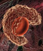
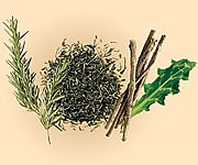
While this is especially true among fair-skinned, blue-eyed people who do not tan well, no one is safe from overexposure to the sun. Even riding in the car with the windows up is no guarantee against sunburn. Studies show that, while automobile and household window glasses screen out UVB rays, they provide inadequate protection from damaging UVA rays,5,6 which is one more reason to include a broad-spectrum sun-screen in your daily health regimen. Skin Cancer: Public Enemy Number OneSkin cancers account for more than 50% of all cancers. Even if one ignores potentially lethal melanomas, non-melanoma skin cancer remains the most common malignancy in humans. In the United States, the incidence of non-melanoma skin cancers—squamous and basal cell carcinomas—is equal to the incidence of malignancies in all other organs combined.7 As noted earlier, sunburn damage is cumulative; while some damage is bad, more damage is even worse. It is never too late to prevent additional damage, and it may be possible to significantly reverse some damage that has already been sustained. This is because recent advances in skin-care science have yielded new approaches to skin repair and restoration. Researchers have discovered botanical extracts that penetrate the outer layers of the skin (epidermis) to reach the dermis, the living layer where skin is constantly repaired and renewed. These extracts have been clinically shown to activate the body’s own repair mechanisms, prompting the reversal of ultraviolet-light-induced damage. Several of these compounds—including phytochemicals derived from green tea, licorice root, milk thistle, and rosemary—have been available to consumers for several years. Now, scientists have identified an exciting new skin-protective agent called beta-glucans. Derived from oats, beta-glucans stimulate the dermal layers of skin to promote remarkable healing and repair from within. Promoting skin collagen synthesisBeta-glucans represent an exciting new development in the “wrinkle wars.” Beta-glucans combined with a collagen matrix are approved by the FDA for use in wound repair among burn victims,8 and beta-glucans have shown great promise in combating the potentially serious tissue damage associated with bedsores.9 It was long believed that the large size of beta-glucans made these bioactive molecules incapable of penetrating the outer layers of intact, erstwhile healthy skin. However, a unique process now allows scientists to extract smaller molecules of beta-glucans from whole grain oats. These “designer” beta-glucans nevertheless retain the immune-stimulating properties of beta-glucans used for oral applications. Moreover, as we shall see, they are adept at promoting collagen synthesis by directly stimulating connective tissue cells within the dermis. Beta-Glucans for Dermal RepairOral supplementation with beta-glucans has been shown to enhance the activity and effectiveness of the body’s immune system.10 Beta-glucans accomplish this by stimulating the activation of macrophages, or white blood cells.11 Macrophages identify, engulf, and destroy bacteria and cancer cells. Macrophages possess specialized receptors that are programmed to recognize and interact with beta-glucans. A given beta-glucan molecule fits into a receptor like a key into a lock, which then “turns on” the macrophage. In the skin, macrophages are enlisted to remove dead cells and help repair wounds. This is where topically applied beta-glucans come in. Tailor-Made Beta-glucansScientists have developed a novel form of beta-glucans that readily penetrate the outer layer of the skin (the stratum corneum), moving down through the epidermis to the living dermal tissue. They believe that these beta-glucans work to benefit the skin in several ways.
First, when applied topically, they form a thin film over the stratum corneum, locking in moisture. Second, they are thought to penetrate deeper layers and circulate in the spaces between live skin cells (keratinocytes) and connective tissue cells (fibroblasts). Here, scientists believe that they stimulate fibroblasts to produce procollagen and collagen, probably by eliciting the release of certain growth factors.12 This reverses some of the undesirable changes in skin associated with aging and cumulative sun damage—changes that are directly related to loss of collagen and procollagen.1 Finally, both systemic and topically applied beta-glucans have been shown to help speed the healing of burn-induced tissue damage, in part by restoring depleted levels of antioxidants.13 It has long been thought that beta-glucans indirectly affect collagen synthesis by stimulating macrophages to release cytokines (proteins that act as cellular mediators), thus stimulating fibroblasts to produce more collagen.9,12,14,15 However, more recent research shows that fibroblasts themselves are studded with specific receptors for beta-glucans.16,17 When glucan molecules bind with these receptors, fibroblasts release proteins known as transcription factors, which initiate genetic transcription. Transcription is the first step in the cellular process that produces new molecules tailored to perform specialized tasks.12,14 Specifically, beta-glucans stimulate fibroblasts to activate genes involved in collagen synthesis18 and to release an array of growth factors that are intimately associated with wound repair and the production of healthy new skin tissue.16 The result is smoother, more youthful-looking skin. Nature’s Arsenal Against PhotoagingBeta-glucans are just one of many natural substances that directly benefit skin. The active ingredients in commercial sunscreens are usually limited to zinc oxide or titanium dioxide to serve as a physical barrier against UV radiation, often in combination with avobenzone or dioxybenzone, which provide approved sun-protection-factor (SPF) ratings.19 There are, however, naturally occurring compounds that protect against light-induced damage and help skin repair and regenerate from within. According to leading photoaging researchers at the University of Alabama at Birmingham, “In recent years, considerable interest has been focused on identifying naturally occurring botanicals, specifically dietary, for the prevention of photocarcinogenesis.”20 Noting that grape seed proanthocyanidins, silymarin (from milk thistle), and green tea polyphenols, among other botanicals, show great promise, they add, “These botanicals may favorably supplement sunscreen protection and may provide additional antiphotocarcinogenic protection, including protection against other skin disorders caused by solar UV radiation.”20 This investigative team recently published a review of research that shows how the above-mentioned botanical compounds act to prevent and even reverse some of the damage associated with UV exposure. Researcher Santosh K. Katiyar de-tails several molecular mechanisms by which these botanical com-pounds protect against cancer, concluding, “The new information regarding the mechanisms of action of these agents supports their poten-tial use as adjuncts in the prevention of [UV-induced cancers].”21
Grape Seed ExtractOne such agent is grape seed extract, or, more specifically, grape seed proanthocyanidins. Chemically speaking, proanthocyanidins are a group of potent antioxidants synthesized by plants as protection from a variety of threats, including UV radiation.22 Grape seed is an excellent source of these natural antioxidants.23
Studies conducted at Ohio State University Medical Center showed that topical application of grape seed proanthocyanidins accelerated the closure and healing of wounded skin in lab rodents.22,24 Noting that wounds treated with grape seed extract demonstrated a variety of indicators of superior healing, the scientists concluded, “Topical application of [grape seed proanthocyanidins] represents a feasible and productive approach to support dermal wound healing.”22 More recently, this same investigative team reported that mice fed grape seed proanthocyanidins had significantly fewer skin tumors after exposure to UVB light than did their counterparts that did not receive supplemental grape seed extract. The few tumors that did appear in supplemented mice were significantly smaller. Grape seed appeared to fight oxidative stress, as mice receiving the extract experienced far less decline in important natural antioxidants, such as glutathione and catalase, than non-supplemented mice. Oxidative stress, in which radiation generates free radicals in the skin, is believed to play a central role in the promotion of skin cancer.25 Dr. Katiyar, who led the study, stated: “These polyphenols have anti-inflammatory and antioxidant properties. Because of these characteristics, polyphenols have been shown to inhibit, reverse, or slow down the risk of UV-induced skin carcinogenesis. As in other organ systems, aging in the skin results in progressive dysfunction. Clinical conditions associated with age-dependent dysfunction include increased ease of wounding, poor wound healing, skin cancer, and infectious disease susceptibility. Clinically, the photoaging component of skin aging accounts for the development in sun-exposed areas of wrinkling, mottled hyperpigmentation and depigmentation, coarsening of the skin, roughness, poor elastic recoil, and bruisability.26 Dr. Katiyar believes natural polyphenols from green tea, grape seed extract, and milk thistle “can inhibit the process of skin aging and photoaging.”26 | ||||||||
Green Tea ExtractGreen tea is possibly one of the world’s oldest sun-protective agents, having been consumed as a “functional food” for at least 4,000 years.27-29 Studies have shown that green tea and green tea extract prevent photoaging both from within (taken orally) and without (when applied topically).30-35 Numerous studies have shown that topically applied green tea extract (or applications of the main bioactive green tea polyphenol, epigallocatechin-3-gallate or EGCG) can prevent the development of skin cancers in lab rodents specially bred for these types of investigations. Mechanisms of action appear to include enhanced DNA repair and retention of robust immune function. Unprotected skin exposed to excessive UV radiation invariably suffers a significant suppression of immune function, a deficit believed to play a role in UV-induced cancers. Furthermore, topical EGCG interferes with tumors’ ability to supply themselves with blood, and stimulates immune system T cells, which function to destroy aberrant cells. Thus, green tea acts to thwart cancer at several stages in its development and spread.36-38 RosemaryRosemary, a perennial herb used as both a culinary and medicinal plant for thousands of years, is packed with a broad array of beneficial compounds, including cancer-fighting chemicals, antioxidants, and anti-inflammatories.39-41 Italian scientists recently identified several rosemary compounds that exhibit potent anti-inflammatory action.41 Two of these compounds—carnosic and ursolic acids—have been shown to be of particular benefit to skin.42-44 Rutgers University researchers showed that topical application of both carnosic and ursolic acids significantly inhibits tumor growth in a mouse model of human skin cancer. Tumor inhibition was as high as 99%, depending on the concentration of the rosemary extract.42 More recently, Indian scientists studied the effects of feeding rosemary extract to specialized lab mice, which serve as surrogate models for human skin cancer. “[The] . . . extract could prolong the latency period of tumor occurrence [and] decrease tumor incidence, tumor burden, and tumor yield,” the researchers concluded.45
Milk Thistle ExtractMilk thistle contains silibinin and silymarin, two flavonoid compounds used for many years to treat liver disease in Europe and Asia. These unique compounds have recently come under increased scrutiny by scientists due to their well-documented antioxidant, anti-inflammatory, and immune-enhancing properties. Numerous studies have shown that these extracts combat skin cancer through a variety of mechanisms, including protecting DNA against damage, decreasing oxidative stress, and lessening inflammation.46-61 Researchers at the University of Michigan noted recently, “Melanoma is one of the few tumors that have increased in incidence over the last few decades.”49 They suggested that silymarin and green tea are among “several promising agents” that could be harnessed to “significantly decrease the morbidity and mortality from this deadly cancer.”49 “Silymarin possesses exceptionally high protective effects against [skin] tumor promotion,” concluded investigators at Case Western Reserve University.53 As such, silymarin is a superb choice for inclusion in sun protection products. It is exceptionally well tolerated and protects against skin cancers through diverse mechanisms. “Silymarin may favorably supplement sunscreen protection and provide additional anti-photocarcinogenic protection,” wrote Dr. Katiyar, in the International Journal of Oncology.46 Licorice Root ExtractLicorice root has been used medicinally since prehistoric times.62 In Chinese medicine, it is one of the oldest and most frequently employed botanicals, with recognized anti-inflammatory, anti-viral, anti-ulcer, and cancer-preventive properties.63 Japanese researchers showed that licorice constituents inhibit the growth of melanoma cancer cells growing in culture. More recently, Japanese scientists demonstrated that a licorice constituent induces a variety of cancer cell types (from liver and stomach cancer to leukemia cells) to undergo apoptosis, or cellular suicide.64 Today, scientists are interested in licorice extract’s ability to promote skin health and avert cancer. Middle Eastern scientists conducted a double-blind study of licorice gel as a treatment for atopic dermatitis, a chronic inflammatory disease of the skin. They concluded, “Licorice extract could be considered as an effective agent for treatment of atopic dermatitis.”65 Italian researchers studying the licorice constituent glycyrrhizin concluded, “Glycyrrhizin treatment might offer protection from the damage induced in humans by UVB radiation.”66 ConclusionMaintaining youthful-looking skin and protecting against cancer require a multipronged approach, one that incorporates both UV blockers and bioactive botanicals for protection from the sun’s aging effects. Simple sunscreens may be inadequate to achieve this level of defense. For total protection and restoration of skin, consider daily use of products that provide scientifically substantiated, natural skin-rejuvenating agents. | |
| References | |
| 1. Quan T, He T, Kang S, Voorhees JJ, Fisher GJ. Solar ultraviolet irradiation reduces collagen in photoaged human skin by blocking transforming growth factor-beta type II receptor/Smad signaling. Am J Pathol. 2004 Sep;165(3):741-51. 2. de Gruijl FR. Skin cancer and solar UV radiation. Eur J Cancer. 1999 Dec;35(14):2003-9. 3. Schober-Flores C. The sun’s damaging effects. Dermatol Nurs. 2001 Aug;13(4):279-86. 4. Gray-Schopfer V, Wellbrock C, Marais R. Melanoma biology and new targeted therapy. Nature. 2007 Feb 22;445(7130):851-7. 5. Hampton PJ, Farr PM, Diffey BL, Lloyd JJ. Implication for photosensitive patients of ultraviolet A exposure in vehicles. Br J Dermatol. 2004 Oct;151(4):873-6. 6. Tuchinda C, Srivannaboon S, Lim HW. Photoprotection by window glass, automobile glass, and sunglasses. J Am Acad Dermatol. 2006 May;54(5):845-54. 7. Miller DL, Weinstock MA. Nonmelanoma skin cancer in the United States: incidence. J Am Acad Dermatol. 1994 May;30(5 Pt 1):774-8. 8. Delatte SJ, Evans J, Hebra A, et al. Effectiveness of beta-glucan collagen for treatment of partial-thickness burns in children. J Pediatr Surg. 2001 Jan;36(1):113-8. 9. Sener G, Sert G, Ozer SA, et al. Pressure ulcer-induced oxidative organ injury is ameliorated by beta-glucan treatment in rats. Int Immunopharmacol. 2006 May;6(5):724-32. 10. Reynolds JA, Kastello MD, Harrington DG, et al. Glucan-induced enhancement of host resistance to selected infectious diseases. Infect Immun. 1980 Oct;30(1):51-7. 11. Demir G, Klein HO, Mandel-Molinas N, Tuzuner N. Beta glucan induces proliferation and activation of monocytes in peripheral blood of patients with advanced breast cancer. Int Immunopharmacol. 2007 Jan;7(1):113-6. 12. Wei D, Zhang L, Williams DL, Browder IW. Glucan stimulates human dermal fibroblast collagen biosynthesis through a nuclear factor-1 dependent mechanism. Wound Repair Regen. 2002 May;10(3):161-8. 13. Toklu HZ, Sener G, Jahovic N, Uslu B, Arbak S, Yegen BC. beta-Glucan protects against burn-induced oxidative organ damage in rats. Int Immunopharmacol. 2006 Feb;6(2):156-69. 14. Wei D, Williams D, Browder W. Activation of AP-1 and SP1 correlates with wound growth factor gene expression in glucan-treated human fibroblasts. Int Immunopharmacol 2002 Jul;2(8):1163-72. 15. Portera CA, Love EJ, Memore L, et al. Effect of macrophage stimulation on collagen biosynthesis in the healing wound. Am Surg. 1997 Feb;63(2):125-31. 16. Kougias P, Wei D, Rice PJ, et al. Normal human fibroblasts express pattern recognition receptors for fungal (1-->3)-beta-D-glucans. Infect Immun. 2001 Jun;69(6):3933-8. 17. Lowe EP, Wei D, Rice PJ, et al. Human vascular endothelial cells express pattern recognition receptors for fungal glucans which stimulates nuclear factor kappaB activation and interleukin 8 production. Winner of the Best Paper Award from the Gold Medal Forum. Am Surg. 2002 Jun;68(6):508-17. 18. Gao CF, Wang H, Wang AH, et al. Transcriptional regulation of human alpha1(I) procollagen gene in dermal fibroblasts. World J Gastroenterol. 2004 May 15;10(10):1447-51. 19. Available at: http://www.fda.gov/cder/otcmonographs/Sunscreen/sunscreen(352).htm. Accessed April 3, 2007. 20. Baliga MS, Katiyar SK. Chemoprevention of photocarcinogenesis by selected dietary botanicals. Photochem Photobiol Sci. 2006 Feb;5(2):243-53. 21. Katiyar SK. UV-induced immune suppression and photocarcinogenesis: Chemoprevention by dietary botanical agents. Cancer Lett. 2007 Mar 21. 22. Khanna S, Venojarvi M, Roy S, et al. Dermal wound healing properties of redox-active grape seed proanthocyanidins. Free Radic Biol Med. 2002 Oct 15;33(8):1089-96. 23. Sen CK, Bagchi D. Regulation of inducible adhesion molecule expression in human endothelial cells by grape seed proanthocyanidin extract. Mol Cell Biochem. 2001 Jan;216(1-2):1-7. 24. Khanna S, Roy S, Bagchi D, Bagchi M, Sen CK. Upregulation of oxidant-induced VEGF expression in cultured keratinocytes by a grape seed proanthocyanidin extract. Free Radic Biol Med. 2001 Jul 1;31(1):38-42. 25. Sharma SD, Meeran SM, Katiyar SK. Dietary grape seed proanthocyanidins inhibit UVB-induced oxidative stress and activation of mitogen-activated protein kinases and nuclear factor-{kappa}B signaling in in vivo SKH-1 hairless mice. Mol Cancer Ther. 2007 Mar;6(3):995-1005. 26. Available at: http://main.uab.edu/uasom/2/show/asp?durki=69362. Accessed March 27, 2007. 27. Weisburger JH. Tea and health: a historical perspective. Cancer Lett. 1997 Mar 19;114(1-2):315-7. 28. Cabrera C, Artacho R, Gimenez R. Beneficial effects of green tea--a review. J Am Coll Nutr. 2006 Apr;25(2):79-99. 29. Yang CS, Prabhu S, Landau J. Prevention of carcinogenesis by tea polyphenols. Drug Metab Rev. 2001 Aug;33(3-4):237-53. 30. Katiyar S, Elmets CA, Katiyar SK. Green tea and skin cancer: photoimmunology, angiogenesis and DNA repair. J Nutr Biochem. 2006 Oct 16. 31. Elmets CA, Singh D, Tubesing K, et al. Cutaneous photoprotection from ultraviolet injury by green tea polyphenols. J Am Acad Dermatol. 2001 Mar;44(3):425-32. 32. Rees JR, Stukel TA, Perry AE, et al. Tea consumption and basal cell and squamous cell skin cancer: Results of a case-control study. J Am Acad Dermatol. 2007 Jan 26. 33. Chiu AE, Chan JL, Kern DG, et al. Double-blinded, placebo-controlled trial of green tea extracts in the clinical and histologic appearance of photoaging skin. Dermatol Surg. 2005 Jul;31(7 Pt 2):855-60. 34. Dvorakova K, Dorr RT, Valcic S, Timmermann B, Alberts DS. Pharmacokinetics of the green tea derivative, EGCG, by the topical route of administration in mouse and human skin. Cancer Chemother Pharmacol. 1999;43(4):331-5. 35. Song XZ, Xia JP, Bi ZG. Effects of (-)-epigallocatechin-3-gallate on expression of matrix metalloproteinase-1 and tissue inhibitor of metalloproteinase-1 in fibroblasts irradiated with ultraviolet A. Chin Med J (Engl). 2004 Dec;117(12):1838-41. 36. Vayalil PK, Elmets CA, Katiyar SK. Treatment of green tea polyphenols in hydrophilic cream prevents UVB-induced oxidation of lipids and proteins, depletion of antioxidant enzymes and phosphorylation of MAPK proteins in SKH-1 hairless mouse skin. Carcinogenesis. 2003 May;24(5):927-36. 37. Chung JH, Han JH, Hwang EJ, et al. Dual mechanisms of green tea extract (EGCG)-induced cell survival in human epidermal keratinocytes. FASEB J. 2003 Oct;17(13):1913-5. 38. Katiyar SK. Skin photoprotection by green tea: antioxidant and immunomodulatory effects. Curr Drug Targets Immune Endocr Metabol Disord. 2003 Sep;3(3):234-42. 39. Ho CT, Wang M, Wei GJ, Huang TC, Huang MT. Chemistry and antioxidative factors in rosemary and sage. Biofactors. 2000;13(1-4):161-6. 40. Calabrese V, Scapagnini G, Catalano C, et al. Biochemical studies of a natural antioxidant isolated from rosemary and its application in cosmetic dermatology. Int J Tissue React. 2000;22(1):5-13. 41. Altinier G, Sosa S, Aquino RP, et al. Characterization of topical antiinflammatory compounds in Rosmarinus officinalis L. J Agric Food Chem. 2007 Mar 7;55(5):1718-23. 42. Huang MT, Ho CT, Wang ZY, et al. Inhibition of skin tumorigenesis by rosemary and its constituents carnosol and ursolic acid. Cancer Res. 1994 Feb 1;54(3):701-8. 43. Offord EA, Gautier JC, Avanti O, et al. Photoprotective potential of lycopene, beta-carotene, vitamin E, vitamin C and carnosic acid in UVA-irradiated human skin fibroblasts. Free Radic Biol Med. 2002 Jun 15;32(12):1293-303. 44. Harmand PO, Duval R, Delage C, Simon A. Ursolic acid induces apoptosis through mitochondrial intrinsic pathway and caspase-3 activation in M4Beu melanoma cells. Int J Cancer. 2005 Mar 10;114(1):1-11. 45. Sancheti G, Goyal PK. Effect of Rosmarinus officinalis in modulating 7,12-dimethylbenz(a)anthracene induced skin tumorigenesis in mice. Phytother Res. 2006 Nov;20(11):981-6. 46. Katiyar SK. Silymarin and skin cancer prevention: anti-inflammatory, antioxidant and immunomodulatory effects (Review). Int J Oncol. 2005 Jan;26(1):169-76. 47. Gazak R, Walterova D, Kren V. Silybin and silymarin--new and emerging applications in medicine. Curr Med Chem. 2007;14(3):315-38. 48. Svobodova A, Zdarilova A, Maliskova J, et al. Attenuation of UVA-induced damage to human keratinocytes by silymarin. J Dermatol Sci. 2007 Apr;46(1):21-30. 49. Lao CD, Demierre MF, Sondak VK. Targeting events in melanoma carcinogenesis for the prevention of melanoma. Expert Rev Anticancer Ther. 2006 Nov;6(11):1559-68. 50. Wright TI, Spencer JM, Flowers FP. Chemoprevention of nonmelanoma skin cancer. J Am Acad Dermatol. 2006 Jun;54(6):933-46. 51. Gu M, Singh RP, Dhanalakshmi S, Mohan S, Agarwal R. Differential effect of silibinin on E2F transcription factors and associated biological events in chronically UVB-exposed skin versus tumors in SKH-1 hairless mice. Mol Cancer Ther. 2006 Aug;5(8):2121-9. 52. Dvorak Z, Vrzal R, Ulrichova J. Silybin and dehydrosilybin inhibit cytochrome P450 1A1 catalytic activity: a study in human keratinocytes and human hepatoma cells. Cell Biol Toxicol. 2006 Mar;22(2):81-90. 53. Lahiri-Chatterjee M, Katiyar SK, Mohan RR, Agarwal R. A flavonoid antioxidant, silymarin, affords exceptionally high protection against tumor promotion in the SENCAR mouse skin tumorigenesis model. Cancer Res. 1999 Feb 1;59(3):622-32. 54. Katiyar SK, Roy AM, Baliga MS. Silymarin induces apoptosis primarily through a p53-dependent pathway involving Bcl-2/Bax, cytochrome c release, and caspase activation. Mol Cancer Ther. 2005 Feb;4(2):207-16. 55. Singh RP, Dhanalakshmi S, Agarwal C, Agarwal R. Silibinin strongly inhibits growth and survival of human endothelial cells via cell cycle arrest and downregulation of survivin, Akt and NF-kappaB: implications for angioprevention and antiangiogenic therapy. Oncogene. 2005 Feb 10;24(7):1188-202. 56. Mallikarjuna G, Dhanalakshmi S, Singh RP, Agarwal C, Agarwal R. Silibinin protects against photocarcinogenesis via modulation of cell cycle regulators, mitogen-activated protein kinases, and Akt signaling. Cancer Res. 2004 Sep 1;64(17):6349-56. 57. Li LH, Wu LJ, Zhou B, et al. Silymarin prevents UV irradiation-induced A375-S2 cell apoptosis. Biol Pharm Bull. 2004 Jul;27(7):1031-6. 58. Dhanalakshmi S, Mallikarjuna GU, Singh RP, Agarwal R. Dual efficacy of silibinin in protecting or enhancing ultraviolet B radiation-caused apoptosis in HaCaT human immortalized keratinocytes. Carcinogenesis. 2004 Jan;25(1):99-106. 59. Singh RP, Agarwal R. Flavonoid antioxidant silymarin and skin cancer. Antioxid Redox Signal. 2002 Aug;4(4):655-63. 60. Singh RP, Tyagi AK, Zhao J, Agarwal R. Silymarin inhibits growth and causes regression of established skin tumors in SENCAR mice via modulation of mitogen-activated protein kinases and induction of apoptosis. Carcinogenesis. 2002 Mar;23(3):499-510. 61. Bhatia N, Agarwal C, Agarwal R. Differential responses of skin cancer-chemopreventive agents silibinin, quercetin, and epigallocatechin 3-gallate on mitogenic signaling and cell cycle regulators in human epidermoid carcinoma A431 cells. Nutr Cancer. 2001;39(2):292-9. 62. Fiore C, Eisenhut M, Ragazzi E, Zanchin G, Armanini D. A history of the therapeutic use of liquorice in Europe. J Ethnopharmacol. 2005 Jul 14;99(3):317-24. 63. Wang ZY, Nixon DW. Licorice and cancer. Nutr Cancer. 2001;39(1):1-11. 64. Hibasami H, Iwase H, Yoshioka K, Takahashi H. Glycyrrhetic acid (a metabolic substance and aglycon of glycyrrhizin) induces apoptosis in human hepatoma, promyelotic leukemia and stomach cancer cells. Int J Mol Med. 2006 Feb;17(2):215-9. 65. Saeedi M, Morteza-Semnani K, Ghoreishi MR. The treatment of atopic dermatitis with licorice gel. J Dermatolog Treat. 2003 Sep;14(3):153-7. 66. Rossi T, Benassi L, Magnoni C, et al. Effects of glycyrrhizin on UVB-irradiated melanoma cells. In Vivo. 2005 Jan;19(1):319-22 |


