Life Extension Magazine®
The year is 2035. As a longtime Life Extension member, you have managed to live in good health until the age of 95. In 2010, you began to benefit from new therapies such as the transplantation of young stem cells into your blood and failing organs. These therapies protected you from killer diseases and premature aging, but were only a partial solution to your problems. Now your biological clock is starting to take its toll. Your cognitive powers are fading, your arteries are beginning to harden, your muscles are weakening, and your energy has begun to ebb. In 2035, it is apparent that major breakthroughs are on the horizon: gene therapies to prevent aging; intelligent nanorobots to repair dysfunctional brain cells; neurostimulatory therapies to regenerate organs and other body parts; young tissues developed from stem cells to rejuvenate life systems; and new technologies to improve vision, hearing, strength, intelligence, sexual prowess, and other attributes. Unfortunately, it is also apparent that it will still take decades to fully implement these advanced breakthroughs, which will be too late for you. The thought has begun to cross your mind that your generation might be the last to face death before a greatly extended healthy life span becomes commonplace. A Life-Saving Voyage into the FutureHowever, all is not lost. Suspended animation, an advanced technology that was perfected in 2030, is readily available at hospitals throughout the world. After you go through this life-saving procedure to stop death in its tracks, you will be cared for at Timeship, a spectacular building on a beautiful site in the US (www.timeship.org).
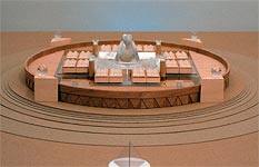
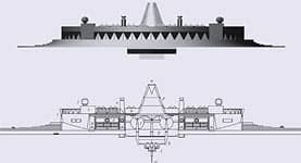
A month later, you are admitted to UCLA Medical Center, where your temperature is gradually lowered to -110 degrees Celsius (-110° C) without ice forming within your body, in a process called vitrification. Before long, you are in a state of biostasis within an insulated capsule. You are ready for a life-saving voyage into the future. Your capsule is then placed into a transport container, which is loaded on to a high-speed, magnetically driven train, which takes you to Timeship, where you will be lowered via an automated system into a geometric honeycomb structure that already contains some of your relatives and friends. (Figs. 1,2.) At Timeship, your body will undergo a stabilization process called annealing, as your temperature is lowered very slowly to -145° C. You will then be cared for with thousands of other patients until the technology has been developed to restore you to health and youthful vigor. Timeship also contains banks of cryopreserved cells, tissues, and organs for use in medicine, as well as laboratories where ongoing research is developing methods to revive and rejuvenate you. Your decades of support for the Life Extension Foundation have clearly paid off. In the 1980s, Life Extension began to fund research to control aging, cure killer diseases, and achieve suspended animation. Life Extension is now funding research at Timeship to restore you to life, health, and youthful vigor. Because of Life Extension, you now have your own very special place in a unique, futuristic ship that is hurtling into time!
This scenario is based on path-breaking research now being funded by the Life Extension Foundation. Life Extension annually funds more than $6 million of research aimed at therapies to extend your life span. We have a visionary plan to make it possible for you to live in good health for centuries. We want to cure you of the ravages of aging and liberate you from the bonds of mortality. In the long run, we are striving for physical immortality. Aging: Our Number-One EnemyAging is the process by which we die. Unless we die in youth, we are subject to the ravages of aging, which depletes our strength, flexibility, cognitive abilities, sexual prowess, coordination, and ability to fight off killer diseases. Even if we manage to avoid heart disease, stroke, cancer, and other killers, we are programmed to lose our health, vitality, and life because of the progressive physical destruction caused by aging. In short, we grow old, suffer, and die. Life Extension’s plan to conquer aging focuses on the study of experimental models of life-span extension. Since aging causes us to die, the best measure of aging is how long we live. In the last 75 years, scientists have found that they can extend life span in a variety of ways. At Life Extension, our primary interest is in methods that extend maximum life span in mammals. Extending Maximum Life SpanThere are currently two experimental models for the extension of maximum life span in mammals: caloric restriction (CR), which has been studied in mice, rats, dogs, and monkeys; and dwarfism in Ames and Snell mice. Calorie-restricted animals are fed a very low-calorie diet enriched with protein, vitamins, minerals, and essential fatty acids, which induces undernutrition without malnutrition. The first studies showing that CR can extend maximum life span were conducted with rats in the 1930s by Clive M. McCay of Cornell University. McCay found that CR could extend the maximum life span of rats by up to 50%, while preventing cancer and other age-related diseases. McCay’s experimental rats were far more youthful and vigorous at advanced ages than rats fed a normal diet.
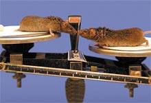
Over the last 75 years, scientists around the world have conducted dozens of CR experiments with similar results. They have found further evidence of CR’s ability to slow aging and extend youth, including its ability to prevent the immune dysfunctions of old age, improve DNA repair, reduce damaging free radical activity, lower glucose and insulin levels, maintain fertility at advanced ages, boost energy levels, reduce the accumulation of damaged proteins, inhibit inflammatory processes, counteract neurodegeneration, and prevent age-related decline in the health-building hormone DHEA (dehydroepiandrosterone). CR also prevents, postpones the incidence of, and reduces the severity of diseases that kill mammals, such as cancer, kidney disease, and cardiovascular disease.1 At advanced ages, calorie-restricted animals are thinner, smaller, and more youthful looking than normally fed animals. The most striking difference between CR and normally fed animals is their activity level. Thirty-month-old, normally fed mice, which are roughly equivalent to 80-year-old humans, are sluggish, bloated, and often have malignant tumors. By contrast, 30-month-old CR mice exercise vigorously, often in acrobatic fashion, and are highly inquisitive. (Fig. 3.) The subjects of CR experiments have included long-lived mammals such as monkeys. In two studies at the University of Wisconsin in Madison and the National Institute on Aging in Baltimore, calorie-restricted monkeys have displayed the youthful characteristics of CR mice and rats, though it is still too early to determine whether their maximum life span will be extended. The other life-span extension model we have been studying is Ames dwarf mice, which have a single gene mutation that interferes with their anterior pituitary function, causing deficiencies in growth hormone, prolactin, and thyroid-stimulating hormone.2 It is believed that the deficiency of growth hormone (and insulin-like growth factor 1, which is made in response to growth hormone) may be the primary reason these mice live so much longer than normal. According to one finding that suggests this, mice that cannot respond to growth hormone because their growth hormone receptor gene has been knocked out live up to 55% longer than normal mice.3 Recently, Dr. Andrzej Bartke and his colleagues at Southern Illinois University showed that Ames dwarf mice, which live 50% longer than normal Ames mice, had their life span extended another 25% by caloric restriction.4 We have been collaborating with Dr. Bartke on studies of gene expression in Ames dwarf mice. Because severe, long-term caloric restriction is not practical in humans, our strategy has been to search for drugs, nutrients, and other therapies capable of extending life span in mammals, or CR mimetics. We quickly concluded that we needed a shortcut to discovering CR mimetics. Life-span studies—even in relatively short-lived species such as mice—simply take too long and are too expensive to make it likely that we can find true CR mimetics in the foreseeable future.
Genes That Extend Maximum Life SpanIn 1999, we came across a new experimental method to help search for CR mimetics: high-density microarrays, or gene chips. Drs. Richard Weindruch and Tomas Prolla of the University of Wisconsin published a study of gene expression in the muscles of both normal and CR mice.5 Gene chips enabled the scientists to rapidly measure changes in the expression of more than 6,000 genes. Not only was this a dramatic shortcut to finding genes involved in extending maximum life span, but it also provided an approach to screening for CR mimetics. Before long, Life Extension was funding gene-chip CR studies at the University of California, Riverside, under the direction of Drs. Stephen R. Spindler and Joseph Dhahbi. Life Extension has now been funding gene-expression research over the past five years, initially at the University of California and then at other laboratories in California and in other states and countries. Most of our funding has been through BioMarker Pharmaceuticals (www.biomarkerinc.com), a company created to discover genetic bio-markers of aging in order to develop anti-aging therapies in humans. Initial Gene-Expression StudiesThe initial gene-expression studies we funded produced provocative results that have been published in scientific journals. In 2003, we decided that we needed to conduct new and larger studies because very few animals were used in the initial studies and because they were conducted with gene chips that could measure the expression of only about 6,000 or 12,000 genes. By 2003, more advanced gene chips were available that could measure the entire mouse genome of 39,000 transcripts, including 34,000 genes. BioMarker established a laboratory in northern California and developed collaborations with Stanford University, Gorilla Genomics, the Chinese Academy of Sciences, and the Children’s Hospital Oakland Research Institute, where we now have access to sophisticated new facilities and greater scientific expertise. Before we look at the gene-expression and other studies Life Extension is now funding, here is a summary of some findings from the initial studies we funded:
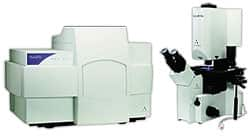
New Research EquipmentAfter our laboratory was launched in northern California in late 2003, a colony of naturally long-lived mice was established for future studies. We purchased young mice and allowed a group of these mice, which had been put on a CR diet, to mature to adulthood. We also purchased or leased new equipment and state-of-the-art informatics and statistical analysis software to help us conduct future studies more effectively. The equipment we obtained includes: 1. The Agilent 2100 Bioanalyzer, a microfluidics-based platform that uses lab-on-a-chip technology for analysis of DNA, RNA, proteins, and cells. (Fig. 4.) 2. The NanoDrop® ND-1000A UV/Vis Spectrophotometer, a highly accurate system designed for extremely small specimens (1-2 microliters). (Fig. 5.) 3. The Arcturus Veritas™ Automated Laser Capture Microdissection and Laser Cutting System, a highly advanced technology that enables us to isolate single or specific types of cells through ultraviolet dissection. (Fig. 6.)
| ||||||||||||||
Metformin Life-Span StudyAfter finding that metformin produces a gene-expression profile similar to that of long-lived CR mice, we decided to study the effects of long-term administration of metformin on mean and maximum life span and other biomarkers of aging in mice. An initial metformin life-span study was begun at the University of California, Riverside, in 2003, but design flaws in this study convinced us to set up another metformin life-span study in our northern California lab. In the new Metformin study, we used a large number of mice fed a normal diet and supplemented with a dose of metformin comparable to that usually used in humans. A control group was fed a normal diet without metformin. This study, begun in early 2004, is expected to be completed around September 2007. In 2004, we repeated high-density microarray studies of the livers of normal and CR mice with new Affymetrix chips that measure the expression of 34,000 genes in order to generate data on the entire mouse genome. We found many changes in gene expression in the CR mice that are different from those found in normal mice. Analysis of these results continues.
Grape Seed Extract with ResveratrolIn September 2004, a group of normally fed mice was supplemented with either synthetic resveratrol or grape seed extract with resveratrol obtained from Life Extension. Our objective was to compare gene expression in the livers of the mice in the grape extract group to gene expression in the livers of normally fed and CR mice. Life Extension’s grape seed extract with resveratrol included two standardized extracts from the seeds and skin of whole red grapes (Vitis vinifera), which contain proanthocyanidins, catechin, epicatechin, gallic acid, and 10% resveratrol from whole red grapes and Polygonum cuspidatum root extract. It also included vitamin C and calcium from calcium ascorbate. When we compared gene expression in the livers of mice fed grape extract with resveratrol to that found in CR animals, we found that substantial numbers of genes were expressed in a similar fashion. These results suggest that the molecular pathways that these genes regulate are activated in both groups, which could mean that the grape seed extract with resveratrol is an anti-aging therapy. Further detailed pathway analysis is under way and we will likely do a life-span study using Life Extension’s grape seed extract in the future. Other Gene-Expression StudiesWe also conducted gene-expression studies of livers from mice treated with two types of the herbal extract cat’s claw. One of the cat’s claw extracts includes oxindole alkaloids (BMcp-1), while the other is depleted of polysaccharides, tannins, and oxindole alkaloids (BMcp-2). Tissues from both groups of mice treated with either BMcp-1 or BMcp-2 were harvested and processed. Specimens have been sent to Stanford University for further gene-chip processing. Image files containing the data from these groups have been received and data analysis is under way. Studies at the Chinese Academy of SciencesIn collaboration with researchers at the Chinese Academy of Sciences in Beijing, Life Extension has funded studies of its grape seed extract with resveratrol, BMcp-1, BMcp-2, and BM-A1 (a plant extract), as well as a green tea and ginkgo biloba extract developed by Dr. Baolu Zhao, an authority in free radical biology. One of the Chinese studies examining the grape seed extract was in Drosophila (fruit flies) that were bred to contain the human mutant alpha-synuclein gene. This gene is expressed in the fruit fly brain similarly to that in humans, so that the flies replicate the essential features of human Parkinson’s disease—age-dependent loss of dopaminergic neurons and motor impairment—that manifests (in the flies) as a progressive loss of climbing ability. Groups of these fruit flies received four doses of the Life Extension grape seed extract in their food. Climbing ability was evaluated in each group and the data were analyzed statistically. The results showed a progressive decline in climbing ability with advancing age, and that this decline was reduced in flies supplemented with grape seed extract, an effect that was more noticeable in male Parkinson’s flies than in female Parkinson’s flies. Interestingly, the dose of grape seed extract that most inhibited the decline in climbing ability of the Parkinson’s flies was similar to that used in a life-span study in which flies given the grape seed extract lived 15% longer than those fed a normal diet. Flies receiving the grape seed extract with natural resveratrol showed greater life-span extension than those receiving synthetic resveratrol. In another study, mitochondria from rat livers were exposed to damaging carcinogens, followed by treatment with either Life Extension grape seed extract or BMcp-2. The results showed that treatment with certain doses of grape seed extract or BMcp-2 protected the mitochondria from damage. Mitchondria are the cellular power plants that fuel the body’s life processes. These findings suggest that Life Extension’s grape seed extract with resveratrol may eventually play a role in preventing or treating Parkinson’s disease in humans. They also suggest that Life Extension’s resveratrol, which is extracted from whole red grapes and Polygonum cuspidatum roots, may be superior to synthetic resveratrol.
Is BM-A1 an Anti-Obesity Therapy?BM-A1, a unique plant extract formula developed by Dr. Zhao of the Chinese Academy of Sciences, contains polyphenols, proanthocyanidins, and flavones derived from green tea and gingko biloba. BM-A1 shows promise for promoting cardiovascular and metabolic health, preventing cancer, and fighting obesity. In one study, the Chinese scientists fed rats a high-fat diet, which caused them to gain weight, develop larger and heavier livers, and demonstrate significant elevations in serum and liver triglyceride levels. Treatment with BM-A1 reduced the weight of rats fed the high-fat diet as well as those fed a normal diet, reduced the size and weight of their livers, and lowered their triglyceride levels. This study identified mechanisms of action involved in preventing obesity, which are linked to an important molecular pathway related to fat metabolism. This pathway involves a set of nuclear receptors known as peroxisome proliferator-activated receptors (PPARs). The scientists found that BM-A1 exerts its anti-obesity effects by modulating PPAR pathways. In doing so, BM-A1 inhibits the accumulation of lipids in adipose tissue, stimulates the utilization of triglycerides, and reduces lipid peroxidation caused by free radical activity. A clinical trial was conducted to gauge BM-A1’s effects on lipid, glucose, and blood pressure levels in humans. The subjects were given BM-A1 and then followed for three months. At the end of this period, 71% showed major lowering of blood lipids and 29% showed moderate lowering; 59% showed major lowering of blood glucose, with 41% showing moderate lowering; and 72% showed major lowering of blood pressure, with 28% showing moderate lowering. A clinical trial examining BM-A1’s effects on obesity in humans needs to be conducted. Since BM-A1’s effects in rats are similar to those produced by CR and metformin in mice, it will be interesting to investigate gene-expression changes in mice given BM-A1. With Life Extension-funded collaborative studies under way at prestigious institutions in the US and China, we expect to move forward with greater speed in identifying the genes and molecular pathways involved in aging and life extension. Using this knowledge, we hope to develop therapies to control aging and extend the healthy human life span. Life-Extending Promise of HypothermiaTerri Schiavo suffered an estimated 10 minutes of cardiac arrest that severely damaged her brain. Afterwards, her relatives fought over whether to keep her in a “vegetative” state on artificial life support (in this case, a feeding tube and pump) or “turn off the machines” and let her die. A court ruling finally settled the matter by deciding that her prognosis was so poor that she should be allowed to die. An autopsy eventually showed that her brain was about half the weight of a normal brain, meaning that about half of its neurons were gone. Every day, people suffer heart attacks that either kill them or cause severe brain damage. This occurs because, at normal body temperature, after about five minutes without oxygenated blood being pumped through the brain, the brain’s neurons suffer lethal damage. Severe brain damage caused by oxygen deprivation may also be caused by other conditions, including strokes and closed head injuries. Although neurons may suffer lethal damage in five to 10 minutes, they do not actually die and disappear until after many hours, or even days. This leaves a window of opportunity to save them. For over 50 years, scientists have known that cooling the brain (and the rest of the body) can have dramatic effects. One of these is to protect against the biochemical processes that kill neurons in brains with inadequate oxygen, a state caused by reduced or absent blood circulation, or ischemia. The success of hypothermia in brain protection is demonstrated in drowning cases, where victims have been revived after as long as 60 minutes of immersion in icy water, because their brains were cooled. This state of cooling is known in medicine as hypothermia. Hypothermia slows down life processes. If applied properly after a person suffers from acute oxygen deprivation, hypothermia has the power to slow down the dying process in damaged neurons. This could save the lives of millions of people. Dogs have been resuscitated after as long as 15 minutes without blood flow at normal body temperature. This was done by cooling the animals with a bypass machine after their hearts were restarted. The same should be able to be done in humans. Patients like Terri Schiavo could be cooled during resuscitation, so that they could eventually live a normal life instead of suffering permanent brain damage. The deepest hypothermia has been achieved in medical centers using a cardiopulmonary bypass system, with a heat exchanger to cool blood directly while it circulates outside the body. Such systems have lowered brain (and body) temperatures rapidly enough to permit doctors to operate successfully on patients in the absence of blood flow. For example, doctors need to induce hypothermia safely in order to perform operations on the vessels of the brain and heart, which cannot be performed while the heart is still beating and blood is flowing. However, induced hypothermia has had limited use in medicine because it has proved difficult to cool patients rapidly in emergencies without inducing some degree of damage. In resuscitation, the use of hypothermia has been limited because it cannot be done rapidly and safely enough in the field to protect the patient completely until he or she can be moved to a well-equipped medical center. Even then, inducing even mild hypothermia takes hours. The vast majority of people who suffer heart attacks, stroke, and head injuries do so at home, work, or play, not when they are in a hospital. In the case of soldiers who suffer cardiac arrest from loss of blood after severe injury, safe and rapid cooling may currently be impossible. Funding Induced Hypothermia ResearchLife Extension has been funding induced hypothermia research at Critical Care Research (CCR), a laboratory in California. Directed by Steven Harris, MD, the CCR research program is developing an advanced technology to deliver safe and rapid cooling in hospitals, clinics, and ambulances. At CCR, scientists have identified several destructive processes that cause damage in oxygen-deprived brains, including an acute inflammatory response that responds to treatment with cold. Cooling the brain after brain injury has proven to be very similar to treating a burn or sports injury with the application of ice. The technology that CCR invented and has been developing is called liquid ventilation lavage. This process involves the use of a fluorocarbon liquid cooled to near-freezing temperature in order to lavage (wash in cycles) the lungs of experimental anesthetized dogs. In this process, the lungs are not completely filled with fluid, and they work as both heat exchangers and gas exchangers. This process is able to produce very rapid cooling of the animal’s brain and body core, from 100° to 85° Fahrenheit (F) in less than 20 minutes. More than 100 dogs have been successfully revived using this process. Most of these animals have shown signs of minor lung damage for a couple of days after revival, but have recovered completely afterwards. Current liquid ventilation lavage research at CCR is aimed at eliminating all lung damage from dogs that undergo rapid cooling, and redesigning the components of the system into a portable unit capable of being operated by paramedics in an ambulance.
Developing Portable Liquid VentilationFor several years, CCR worked with an engineering firm to place its lavage equipment into a prototype portable system. This required a redesign and computerization of the system’s first pump module, which originally weighed more than 100 lbs. The design eventually was reduced to a double-chamber piston system, which weighed less than 35 lbs. and was small enough to be carried in one hand with a strap. This system needed to be gas powered from a small pressure tank, and also required a vacuum source. Another CCR invention is portable heat exchangers made from pre-frozen “ice cartridges,” which work by routing fluorocarbon through fine tubes immersed in ice. With this system, the amount of ice needed for adequate cooling of a 45-lb. dog was reduced to only 8 lbs. CCR continues to conduct dog experiments with this system. (Fig. 7.) In 2003, CCR engaged the services of a second engineering team to see whether it could help solve some problems that had developed with the first portable system. The new team proposed a redesign of the system that would use only electrically driven pumps. This allowed for direct electrical drive of both lavage liquid infusion and suction, and eliminated the need for both the vacuum and compressed air sources that drive the older system. A new prototype lavage machine has now been built as a breadboard device and has been tested successfully in animal experiments. A few problems remain. Future devices will address problems in the delicate timing of lavage cycles under software control. This approach is expected to result in a smaller, more portable system driven directly by AC (alternating current) power or batteries, which will do away with the need for a heavy vacuum or compressor system. Critical Care Research now routinely cools dogs from 100° to 92° F by liquid lavage with almost no lung problems, resulting in animals that are up and walking the next day, usually “without even a wheeze,” according to Dr. Harris. He notes that since CCR published the first report of fluorocarbon lung lavage cooling in 2001, the lavage method for cooling and heating animals has been confirmed in other laboratories working with rabbits and pigs. With liquid lavage, there is not as much liquid to move compared with total liquid ventilation, and so there is less pressure and damage. Gas ventilation can be used simultaneously to take care of oxygen and carbon dioxide, so that the animal can be cooled without damage. | ||||
Suspended Animation ResearchA related research project under development at CCR is a liquid ventilation system for ultra-rapid cooling. For most medical uses, only moderate-to-rapid cooling for a relatively short period is needed. To develop the first phase of suspended animation, however, more rapid cooling is necessary because the objective is to continue cooling the patient until long-term care can be provided at ultra-low temperatures. CCR has demonstrated that a larger heat exchanger can facilitate an adequate cooling rate for this purpose. Life Extension is also funding the design and construction of a computerized perfusion system at a laboratory in Florida for use in suspended animation research, as well as a new advanced cryogenic system at a laboratory in New York to provide safe, stable, long-term care for cryopreserved humans. Long-Term Suspended AnimationThe key to long-term suspended animation is cooling patients to a temperature low enough to stop all biological processes indefinitely without damaging the patient. The technology needed to accomplish this is known as vitrification. Life Extension funds vitrification research at another laboratory in California called 21st Century Medicine, or 21CM (www.21CM.com). Scientists at 21CM are making rapid progress in developing vitrification methods to cryopreserve cells, tissues, organs, and entire organisms. In the process of striving for its ultimate goal—achieving long-term suspended animation in humans—21CM is making major advances to help the development of medical fields such as transplantation, regeneration, and rejuvenation. 21CM’s vitrification technology will be needed to create banks of cryopreserved tissues and body parts for use in therapies to cure killer diseases and make people younger in the future. To achieve these goals, Life Extension is funding several vitrification research projects at the 21CM lab.
Transplanting Cryopreserved KidneysDr. Gregory Fahy, 21CM’s chief scientific officer, is the world’s foremost expert in the vitrification of biological systems. Dr. Fahy has been working for decades to cryo-preserve kidneys well enough to transplant them successfully, first at the American Red Cross in Maryland and now at 21CM in California. The 21CM lab is the only one in the world currently pursuing vitrification of large tissue masses. Another key scientist working at this lab is Dr. Brian Wowk, who has developed the world’s first practical ice-blocking compounds, which are used with combinations of cryoprotective chemicals in 21CM’s vitrification solutions. These solutions allow tissues to be cooled to extremely low temperatures without the formation of ice. At about -123° C, these tissues are transformed into a glass-like state, which differs dramatically from the crystalline state that occurs during freezing. Vitrification stops life processes cold, without the structural and biochemical damage caused by ice crystals. (Fig. 8.) Survival of the First Vitrified KidneyAt the annual meeting of the Society for Cryobiology in July 2005, Dr. Fahy announced the survival of a rabbit that received a transplanted kidney after the organ had been vitrified to -130° C and then re-warmed. This result was based on the vitrified kidney’s ability to provide sole renal life support in the recipient animal until it was removed for examination 48 days after it was transplanted. The transplanted kidney sustained some damage from ice formation in its center, but that damage was entirely absent in other parts of the core of the kidney, enabling the organ to support life in a normal fashion. This remarkable achievement is a major medical milestone that moves us closer to establishing banks for the long-term cryopreservation of human organs. This research is currently being written up for publication in a scientific journal, while further research to perfect the vitrification of rabbit kidneys continues. In 2004, Dr. Fahy and colleagues published a paper reporting the survival of eight rabbits that had received re-warmed transplanted kidneys that had been cooled to -45° C. This breakthrough was based on earlier advances involving the ability to control cryoprotectant toxicity, nucleation, ice crystal growth, and chilling injury.10 21CM has also had success in developing new methods for short-term storage of kidneys and hearts, as well as vitrification of cartilage and corneas. It has been particularly successful in vitrifying corneas, which cannot be frozen effectively. Corneas are complex structures in the eye that often become opaque with advancing age, which can result in blindness. Corneas vitrified by 21CM scientists generally show no cell loss or cell death, and remain transparent after transplantation. Vitrifying Brains and Whole OrganismsThe most ambitious research being performed at the 21CM laboratory involves experiments to vitrify rabbit brain slices, entire rabbit brains, and entire rabbits. The short-term potential of this research includes the use of well-preserved brain slices to test potentially therapeutic drugs and antidotes to potential biological and chemical weapons of mass destruction. This research could also lead to the availability of well-preserved brain sections for neurobiology experiments and the treatment of brain diseases such as Parkinson’s and Alzheimer’s. This research is also essential for achieving suspended animation in humans, which could be instrumental in radically extending the life span of millions of people.
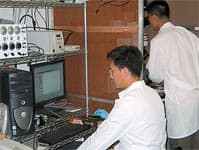
Recovering Viability in Brain SlicesOne test we have used to evaluate brain slice function after vitrification and re-warming is the potassium/ sodium ratio, or ion transport capacity. Another is to determine whether there is normal preservation of slice structure. These tests have shown apparently normal structure and function in hippocampal brain slices. The hippocampus is an area of the brain involved in memory consolidation, storage, and retrieval, and is the part of the brain most sensitive to oxygen deprivation. To further evaluate the effectiveness of its brain studies, 21CM has established an in-house neurophysiology laboratory to measure electrical activity in brain slices and sections. This lab is headed by Dr. Yuansheng Tan, who is assisted by Joon Chang. (Fig. 9.) In initial studies, it was determined that vitrified, re-warmed brain slices were alive but electrically silent. After several refinements in technique, the 21CM brain slice team was able to achieve essentially normal electrical responses in up to seven of nine slices, which compares favorably to the rate of normal electrical responses in uncooled control slices not exposed to vitrification solutions, and exceeds the rate of electrical responses in uncooled control slices in many other laboratories. This is the first time electrical activity analogous to a normal electroencephalogram (EEG) response has been achieved in organized brain tissue after cooling to temperatures low enough to achieve vitrification. Vitrifying the Entire BrainLike studies of isolated hippocampal slices, studies of entire rabbit brains have found that all regions of the brain appear to be structurally preserved after vitrification and re-warming. Initial studies have shown that it is difficult to preserve the brain’s ability to respond electrically after five hours of cold storage or cold perfusion in the absence of cryoprotectant chemicals. Studies are under way to overcome this problem, however, and the 21CM team will eventually look at electrical activity in brains that have been perfused with vitrifiable concentrations of cryoprotectants. It appears as though the entire brain can be vitrified, but further research is required to see whether normal electrical activity in the brain can be preserved after vitrification. Vitrifying the Entire BodyWhen whole rabbits were vitrified, scientists collected tissue samples from many brain and kidney regions as well as from the heart, lungs, liver, intestine, muscle, skin, fat, stomach, and other areas. Using a device called a differential scanning calorimeter, scientists tested the samples to see whether ice formed within them. The differential scanning calorimeter can cool tissue samples to vitrifiable temperatures, warm them up, and measure how much ice has melted during warming. The amount of melted ice is equal to the total amount of ice formed during both cooling and warming, and the temperature at which the ice melts is a measure of how much cryoprotectant was in the tissue before it was cooled. The results showed that the most difficult tissue to vitrify in the entire body was the center of the kidney, called the inner medulla. Most of the tissue samples showed only small amounts of ice formation, or none at all. Since 21CM is moving toward successful vitrification of whole kidneys, it appears that the entire body can be successfully vitrified as well. This suggests that whole-body suspended animation is an achievable goal, but a great deal of additional research will be necessary to attain this goal. Living in Good Health for CenturiesBefore suspended animation is achieved, the vitrification research funded by Life Extension will lead to banks of well-preserved cells, tissues, and organs for use in transplantation, regeneration, and rejuvenation therapies. One obstacle to the development of these life-extending therapies is that transplanted tissues are currently rejected by the body’s immune system, which requires the lifelong administration of expensive immunosuppressive drugs that have serious side effects. This is why transplantation today is largely reserved for patients whose lives are at stake unless they receive a new heart, liver, or kidney. However, research in neutralizing the body’s rejection of foreign tissue is now at an advanced stage, and cloning and stem cell research to develop tissues that are not rejected at all is also advancing rapidly. Overcoming the rejection problem and perfecting organ vitrification will mark the dawning of a new era in which the widespread availability of banked, vitrified body parts will be used to keep us alive, healthy, and vigorous . . . perhaps for centuries! In the scenario that opened this article, you had been placed in suspended animation in 2035 to await the time when advances in science and technology would enable doctors to restore you to life, health, and youth. Based on research now being funded by Life Extension and others, it is possible that you could be revived in a state of physical immortality well before the end of the twenty-first century. | ||||
| References | ||||
| 1. Weindruch R, Walford RL. The Retardation of Aging and Disease by Dietary Restriction. Springfield, IL: Charles C. Thomas Publisher, Ltd; 1988. 2. Bartke A. Genetic models in the study of anterior pituitary hormones In: Shire JGM, ed. Genetic Variation in Hormone Systems. Boca Raton, FL: CRC Press; 1979:113-26. 3. Coschigano KT, Clemmons D, Bellush LL, Kopchick JJ. Assessment of growth parameters and life span of GHR/BP gene-disrupted mice. Endocrinology. 2000 Jul;141(7):2608-13. 4. Bartke A, Wright JC, Mattison JA, et al. Extending the life span of long-lived mice. Nature. 2001 Nov 22;414(6862):412. 5. Lee CK, Klopp RG, Weindruch R, Prolla TA. Gene expression profile of aging and its retardation by caloric restriction. Science. 1999 Aug 27;285(5432):1390-3. 6. Dhahbi JM, Kim HJ, Mote PL, Beaver RJ, Spindler SR. Temporal linkage between the phenotypic and genomic responses to caloric restriction. Proc Natl Acad Sci USA. 2004 Apr 13;101(15):5524-9. 7. Spindler SR. Rapid and reversible induction of the longevity, anticancer and genomic effects of caloric restriction. Mech Ageing Dev. 2005 Sep;126(9):960-6. 8. Cao SX, Dhahbi JM, Mote PL, Spindler SR. Genomic profiling of short- and long-term caloric restriction effects in the liver of aging mice. Proc Natl Acad Sci USA. 2001 Sep 11;98(19):10630-5. 9. Dhahbi JM, Mote PL, Fahy GM, Spindler SR. Identification of potential caloric restriction mimetics by microarray profiling. Physiol Genomics. 2005 Sep 27. 10. Fahy GM, Wowk B, Wu J, et al. Cryopreservation of organs by vitrification: perspectives and recent advances. Cryobiology. 2004 Apr;48(2):157-78. |

