What's Hot
What's Hot
| News flashes are posted here frequently to keep you up-to-date with the latest advances in health and longevity. We have an unparalleled track record of breaking stories about life extension advances.
Meta-analysis confirms protective benefit for magnesium against stroke
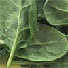
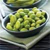
"To our knowledge, the epidemiologic evidence on the relation between dietary magnesium intake and risk of stroke has not yet been summarized," Susanna C. Larsson and her associates at Sweden's Karolinska Institutet write. "Therefore, we performed a systematic review and dose-response meta-analysis to assess the association between magnesium intake and risk of total stroke and stroke subtypes." For their review, Dr Larsson and colleagues selected seven prospective studies that included a total of 241,378 participants and 6,477 stroke cases. Their analysis found an 8 percent reduction in the risk of total stroke in association with each 100 milligram daily increase of magnesium, which remained unchanged following adjustment for the presence of hypertension and diabetes. When strokes were examined according to type, the protective effect of magnesium was confirmed for ischemic stroke, but not intracerebral or subarachnoid hemorrhage, however, the authors note that the low number of hemorrhagic strokes that occurred among the study populations reduced the ability to accurately estimate their association with the mineral. Protective mechanisms for magnesium posited by Dr Larsson and associates include an ability to reduce blood pressure, glucose, lipids, and lipoprotein peroxidation, as well as possibly lowering the risk of type 2 diabetes. The authors recommend increased consumption of magnesium-rich foods, such as leafy vegetables, beans and whole grains as a possible means of decreasing stroke risk. —D Dye Scientists identify molecule's role in calorie restriction brain benefit
Giovambattista Pani, of the Institute of General Pathology, Faculty of Medicine at the Catholic University of Sacred Heart in Rome and his associates found that calorie restriction's beneficial effects on neuronal plasticity, memory and social behavior do not occur in mice lacking a molecule known as cAMP responsive-element binding (CREB)-1 in their forebrains. Calorie restricted animals that are deficient in the molecule showed reduced expression of the sirtuin Sirt-1, as well as a decrease in the induction of genes involved in the metabolism and survival of neurons. The team's finding is consistent with what is known concerning CREB1 in regard to its ability to regulate brain functions such as memory and learning, as well as the decline in its activity that occurs with aging. "Thus, our findings identify for the first time an important mediator of the effects of diet on the brain," Dr Pani stated. "This discovery has important implications to develop future therapies to keep our brain young and prevent brain degeneration and the aging process. In addition, our study shed light on the correlation among metabolic diseases as diabetes and obesity and the decline in cognitive activities." "Our hope is to find a way to activate CREB1, for example through new drugs, so to keep the brain young without the need of a strict diet," he added. —D Dye EPA derivative cures leukemia in mice
In recent research, cyclooxygenase-derived cyclopentenone prostaglandins (CyPGs) were identified as possible agents to target cancer stem cells. Currently available treatments for leukemia and other cancers fail to destroy stem cells, which results in relapses of the disease. "The patients must take the drugs continuously," noted study coauthor Robert F. Paulson, who is an associate professor of veterinary and biomedical sciences at Penn State. "If they stop, the disease relapses because the leukemia stem cells are resistant to the drugs." For the current experiments, the researchers administered a CyPG compound known as delta-12-protaglandin J3 (D12-PGJ3, derived from EPA) to leukemic mice for one week. Animals that received the compound had normal spleens and blood counts, and increased survival without relapse after being treated. "This treatment completely eradicated leukemia stem cells in vivo, as demonstrated by the inability of donor cells from treated mice to cause leukemia in secondary transplantations," the authors write. "Research in the past on fatty acids has shown the health benefits of fatty acids on cardiovascular system and brain development, particularly in infants, but we have shown that some metabolites of omega 3 have the ability to selectively kill the leukemia-causing stem cells in mice," stated Dr Prabhu, who is an associate professor of immunology and molecular toxicology at Penn State's Department of Veterinary and Medical Sciences. "The important thing is that the mice were completely cured of leukemia with no relapse." —D Dye Higher selenium, nickel levels associated with protection against pancreatic cancer
A team from the Spanish National Cancer Research Center and the University of Barcelona conducted a case-control study involving 118 men and women diagnosed with exocrine pancreatic cancer and 399 cancer-free control subjects residing in Spain. Toenail samples were analyzed for arsenic, cadmium, lead, nickel, selenium and other trace elements. Having a high level of arsenic was associated with double the adjusted risk of exocrine pancreatic cancer than the risk experienced by those with a low level, and having high levels of cadmium and lead were associated with a 3.5 and six times greater risk. Subjects with a high level of selenium had a 95 percent lower risk of the disease, and those with high levels of nickel had a 73 percent decrease. While protective benefits for selenium have been indicated by numerous studies, occupational exposure to nickel has been associated with an increased risk of some cancers, however, the researchers remark that nickel may be associated with higher amounts of potentially carcinogenic polychlorinated biphenyls in occupational settings, which could account for the elevated cancer risk. "Aberrant expression patterns of some selenoproteins show that they are relevant in scavenging reactive oxygen species and diminishing oxidative damage," Andre F. S. Amaral and his colleagues write. "Selenium seems also to play a role as an antagonist of arsenic, cadmium and lead, decreasing the oxidative stress caused by exposure to these elements." They conclude that the findings point to a role of trace elements in the development of cancer of the pancreas and warrant further research. —D Dye Soy isoflavones enhance cancer radiation therapy
In earlier research published in the September, 2010 issue of Nutrition and Cancer, Dr Hillman's team found that prostate cancer patients undergoing radiation therapy who consumed soy isoflavones had less toxicity to surrounding tissue and fewer associated side effects such as incontinence compared to untreated subjects. The compounds increase the effect of radiation in cancerous cells by inhibiting DNA repair mechanisms and molecular survival pathways activated by radiation that are not activated in normal cells. Soy isoflavones simultaneously reduce radiation damage in surrounding, healthy tissue. The current research confirmed a benefit for soy isoflavones in cultured non-small cell lung cancer cells and in non-small cell lung tumors in mice treated with radiation. "If we succeed in addressing preclinical issues in the mouse lung cancer model showing the benefits of this combined treatment, we could design clinical protocols for non-small cell lung cancer to improve the radiotherapy of lung cancer," stated Dr Hillman, who is a professor of radiation oncology at Wayne State University's School of Medicine and the Barbara Ann Karmanos Cancer Institute. "We also could improve the secondary effects of radiation, for example, improving the level of breathing in the lungs." "In contrast to drugs, soy is very, very safe," she added. "It's also readily available, and it's cheap. The excitement here is that if we can protect the normal tissue from radiation effects and improve the quality of life for patients who receive radiation therapy, we will have achieved an important goal." —D Dye High parathyroid, low vitamin D levels associated with greater risk of sudden cardiac death
"Disturbances in calcium-phosphorus metabolism, 25-hydroxyvitamin D, and parathyroid hormone (PTH) have been implicated as novel risk factors for cardiovascular events," the authors write. "Disruption of the vitamin D receptor stimulates the renin-angiotensin system, provoking vasoconstriction, oxidative stress, and left ventricular hypertrophy. Parathyroid hormone excess increases intracellular calcium and increases protein synthesis and total cell mass within cardiac myocytes. These mechanisms also contribute to the pathogenesis of sudden cardiac death (SCD), but associations of vitamin D deficiency and PTH with SCD have not been reported." The study evaluated 2,312 men and women who were free of cardiovascular disease upon enrollment in the Cardiovascular Health Study, which studied the risk of the disease in older individuals. Blood samples stored between 1992 and 1993 were analyzed for serum 25-hydroxyvitamin D and parathyroid hormone levels. Participants were followed for a median of 14 years, during which 73 sudden cardiac deaths occurred. While only two sudden cardiac deaths per 1000 occurred among those whose 25-hydroxyvitamin D levels were at least 20 nanograms per milliliter, the amount doubled in those whose levels were lower. Similarly, high parathyroid levels of 65 picograms per milliliter or more were associated with twice the number of SCDs compared to lower levels. For the 11.7 percent who had low vitamin D and high parathyroid levels, the risk of sudden cardiac death was more than twice that of subjects with normal levels. The authors conclude that low vitamin D and high parathyroid hormone levels are independently associated with SCD risk in men and women without cardiovascular disease and recommend further studies. —D Dye Inflammation lowers critical nutrients
A team at the University of Glasgow in Scotland evaluated data from 1,303 men and women whose blood samples underwent testing for C-reactive protein (CRP, a marker of inflammation) as well as micronutrient analysis for vitamins A, B6, C and E, copper, selenium and zinc. Vitamin D levels measured in 3,676 subjects were also included in the study. As C-reactive protein level rose, micronutrient concentrations declined, with the exceptions of vitamin E and copper. A reduction in vitamins B6 and C was observed in association with only slightly increased CRP levels of 5 to 10 milligrams per liter (mg/L). When CRP levels exceeded 80 mg/L, the percentage of subjects with micronutrient levels lower than the reference limit increased by 41% for vitamin A, 53% for vitamin B-6, 47% for vitamin C, 19% for vitamin D and 27% for selenium compared to the percentage associated with CRP levels of 5 mg/L or less. The authors conclude that "zinc is interpretable when CRP concentrations are less than 20 mg/L, plasma selenium and vitamins A and D are interpretable when CRP concentrations are less than 10 mg/L, and vitamins C and B6 can only be interpreted when CRP concentrations are less than 5 mg/L." —D Dye Cherry juice improves sleep
Glyn Howatson and colleagues from Northumbria University compared the effects of Montmorency tart cherry juice or a placebo drink in 20 men and women aged 18 to 40 years. Participants were instructed to consume one serving upon awakening and another before bed for seven days. The subjects subsequently switched regimens following a two-week period in which no drinks were administered. Daily diaries recorded information on the participants' sleep quality, which was corroborated with a wearable sleep monitor. Urine samples obtained before and during the trial were analyzed for 6-sulphatoxymelatonin, the major metabolite of melatonin: a sleep-promoting hormone that occurs in cherries. Participants who received tart cherry juice had significant elevations in urinary melatonin content, while the placebo group's levels remained the same as those measured at the beginning of the trial. Drinking cherry juice was associated with an average of 39 minutes longer sleep, more time in bed spent asleep, better sleep quality and less daytime napping compared to the placebo group. While increased melatonin is the primary mechanism to which the improved sleep of those who consumed cherry juice was attributed, the authors note that sleep regulation is also influenced by pro-inflammatory cytokines and that tart cherries have numerous phenolic compounds that have anti-inflammatory properties. "This is the first study to show direct evidence that dietary supplementation with a tart Montmorency cherry juice concentrate increases circulating melatonin and can provide modest improvements in sleep time and quality in healthy adults with no reported disturbed sleep," they announce. "Tart cherry juice concentrate might therefore present a suitable adjunct intervention for disturbed sleep across a number of scenarios in healthy and symptomatic individuals." —D Dye Aspirin may be okay before heart surgery
Jianzhong Sun and colleagues studied the effect of aspirin on the outcome of 4,256 patients at the University of California Davis Medical Center and Thomas Jefferson University Hospital who underwent coronary artery bypass graft surgery or valve surgery between 2011 and 2009. One thousand nine hundred twenty-three subjects reported consuming aspirin at least once within five days of their surgery, in comparison with 945 who reported no aspirin use. In the month following their surgeries, aspirin users experienced 34 percent fewer major events, including a 61.6 percent lower risk of renal failure, a 56 percent lower risk of needing dialysis and a 39 percent lower risk of premature mortality compared to those who did not use aspirin. Those who used aspirin also spent less time in intensive care. "We know that aspirin can be lifesaving for patients who have experienced heart attacks," stated coauthor Nilas Young, who is chief of cardiothoracic surgery at the University of California Davis Medical Center. "Now we know that this simple intervention can do the same for patients who undergo certain coronary surgeries. This outcome could lead to new preoperative treatment standards in cardiac medicine." Thomas Jefferson University Hospital anesthesiology chair Zvi Grunwald added that "While we are excited that the study clearly showed that preoperative use of aspirin significantly reduced major complications and mortality in patients undergoing cardiac surgery, we do urge further study before recommending aspirin for cardiac surgery patients prior to surgery." —D Dye Green tea compound could help protect transplanted liver against hepatitis C virus reinfection
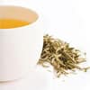
"Green tea catechins such as EGCG and its derivatives epigallocatechin (EGC), epicatechin gallate (ECG), and epicatechin (EC) have been shown to exhibit antiviral and antioncogenic properties," noted Dr Ciesek, who is affiliated with Hannover Medical School's Department of Gastroenterology, Hepatology and Endocrinology. "Our study further explores the potential effect these flavonoids have in preventing HCV reinfection following liver transplantation." The researchers found that while EGCG had no effect on HCV RNA replication, the compound inhibited entry into cultured human liver cells as well as liver tumor cells. Although pretreatment of cells with EGCG before inoculation with HCV failed to reduce infection, applying it during inoculation resulted in significant inhibition. The team also demonstrated that the compound prevented the initial step in infection of a cell by HCV, known as viral attachment. "The green tea antioxidant EGCG inhibits HCV cell entry by blocking viral attachment and may offer a new approach to prevent HCV infection, particularly reinfection following liver transplantation." Dr Ciesek concluded. —D Dye Review confirms glucose reduction benefit for cinnamon in diabetics
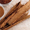
Drs Davis and Yokoyama selected 8 randomized, placebo-controlled trials of cinnamon and/or cinnamon extract in patients with diabetes or prediabetes for their review. Three trials were new and five were included in previous meta-analyses. While the intake of cinnamon or cinnamon extract was associated in a decrease in fasting blood glucose, analysis of cinnamon extract alone also confirmed a significant reduction. Water extracts of cinnamon contain concentrated amounts of compounds known as procyanidins, which are believed to be the ingredients responsible for lowering glucose levels. "The fact that water extracts of cinnamon have (bioactive) activity suggests that these may be preferable in terms of use compared with whole cinnamon," the researchers stated. "Using water extracts of cinnamon achieves the desired blood glucose effects while avoiding the nonpolar constituents in whole cinnamon or the cinnamon flavor components that have been linked to deleterious effects (e.g., oral lesions and mutagenicity)." Drs Davis and Yokoyama concluded that the analysis' results "show that consuming cinnamon, especially cinnamon extract, does produce a modest but statistically significant lowering in fasting blood glucose." —D Dye |

 December 30, 2011. The results of a meta-analysis reported in an article published online on December 28, 2011 in the
  December 28, 2011. In an article published online on December 21, 2011 in the
  December 23, 2011. Sandeep Prabhu and colleagues at Pennsylvania State University report in the December 22, 2011 issue of the journal
  December 21, 2011. A report published online on December 20, 2011 in the journal
  December 19, 2011. While a concern has been expressed that the consumption of beneficial nutritional compounds such as soy isoflavones might protect cancerous tumors from destruction by therapeutic ionizing radiation, in the November, 2011 issue of
  December 14, 2011. In the December, 2011 issue of
  December 12, 2011. Research described online on December 7, 2011 in the
  December 9, 2011. An article published on November 8, 2011 in the
  December 7, 2011. While patients facing surgery are generally recommended to avoid aspirin due to the risk of increased bleeding, a study appearing online on October 12, 2011 in the
  December 5, 2011. The December, 2011 issue of the journal
  December 2, 2011. The results of a meta-analysis published in the September, 2011 issue of the